Quick Facts
Sympathetic Contribution: Spinal cord levels T10-T11.
Parasympathetic Contribution: Vagus nerve.
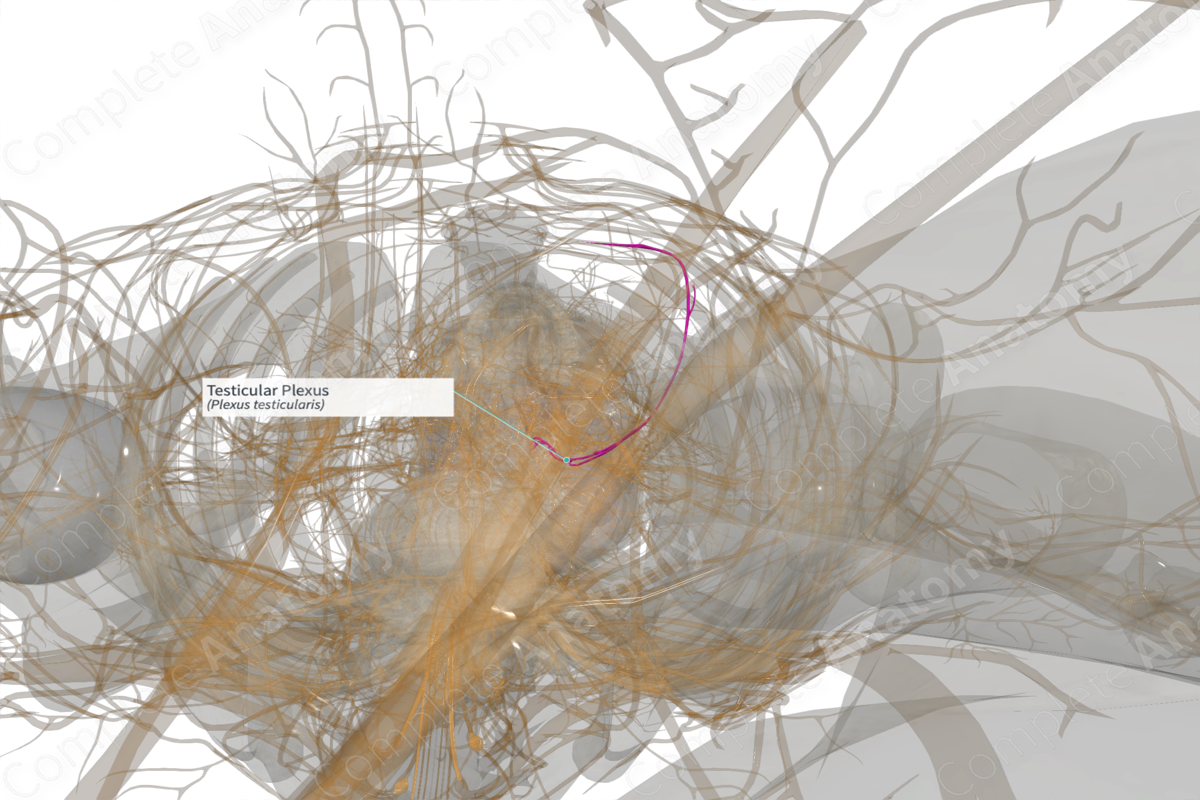
Course: Follows the testicular artery to the testis.
Sympathetic Supply: Testis.
Parasympathetic Supply: Testis.
Contributing Nerves
The testicular plexus is derived from axons that emerge from the aortic plexus and follow the testicular arteries. Sympathetic axons originate at the T10-T11 spinal cord levels, while parasympathetic contributions are vagal. Autonomic fibers traveling through the renal and intermesenteric plexuses will form the testicular plexus (Standring, 2016; Moore, Dalley and Agur, 2013).
Course
The testicular plexus is a periarterial plexus. In this case it is located by the origin of the testicular artery. Its axons travel along the testicular artery to the testis.
Branches
There are no named branches.
Supplied Structures
The testicular plexus supplies sympathetic and parasympathetic axons to the testis and returns visceral sensory information to the central nervous system. The testis may also receive innervation from the inferior hypogastric plexus (Standring, 2016).
References
Moore, K. L., Dalley, A. F. and Agur, A. M. R. (2013) Clinically Oriented Anatomy. Clinically Oriented Anatomy 7th edn.: Wolters Kluwer Health/Lippincott Williams & Wilkins.
Standring, S. (2016) Gray's Anatomy: The Anatomical Basis of Clinical Practice. Gray's Anatomy Series 41 edn.: Elsevier Limited.
Learn more about this topic from other Elsevier products
Plexus

Visceral plexuses are a network of nerve fiber and ganglia surrounding organs of the abdomen and pelvis region that convey sympathetic, parasympathetic, and visceral afferent input.


