Quick Facts
Sympathetic Contribution: T10-L1 spinal cord level.
Parasympathetic Contribution: S2-S4 spinal cord level.
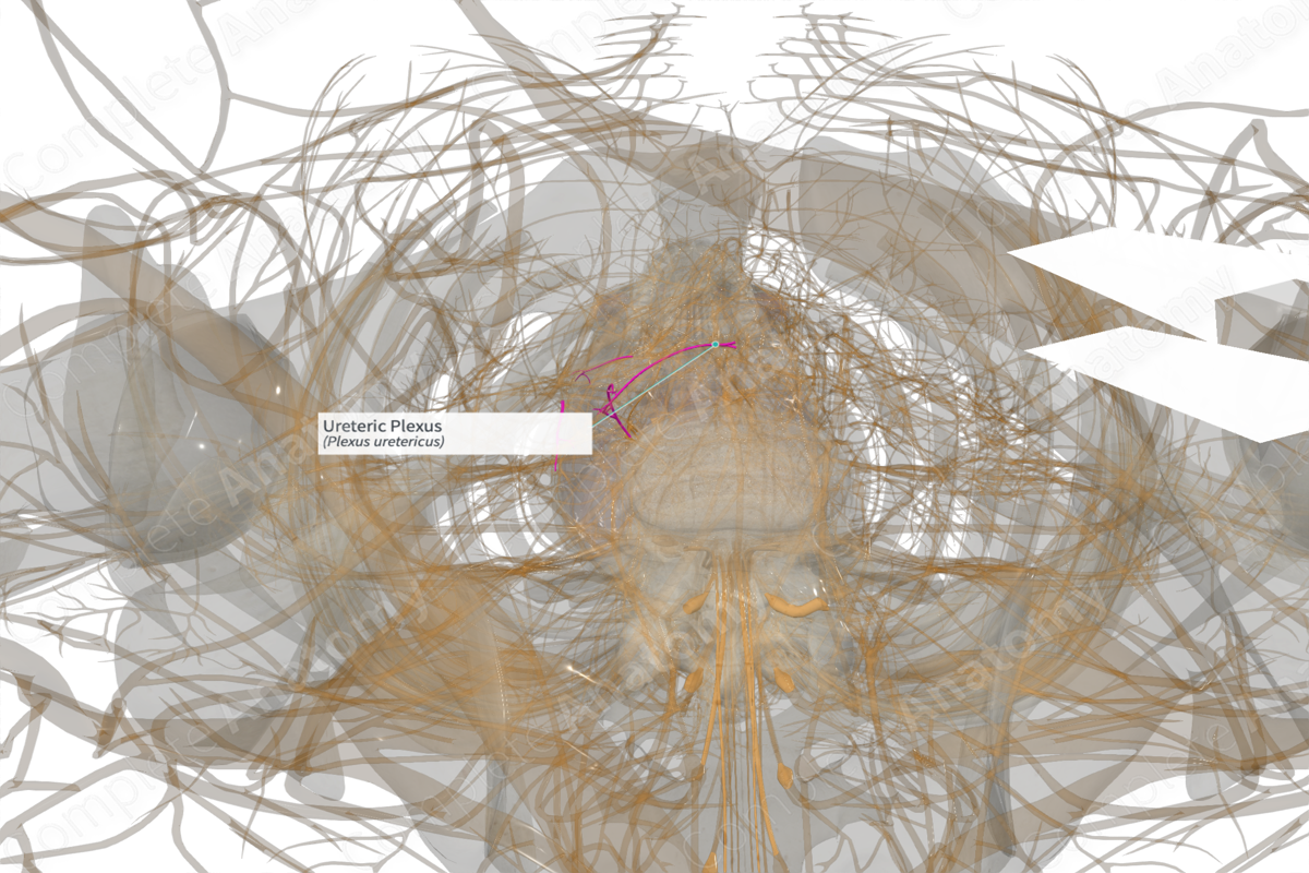
Course: Ureteric plexus runs along the length of the ureters.
Sympathetic Supply: The vasculature and smooth muscle of the ureters.
Parasympathetic Supply: Smooth muscle of the ureters.


Contributing Nerves
The efferent fibers targeting the ureteric plexus originate at the T10-L2 levels for sympathetic and S2-S4 levels for parasympathetic fibers.
The plexus itself is thought of in three sections. The upper section serving the ureter closest to the kidney is derived from the renal and aortic plexuses. The middle section of the ureteric plexus serving the middle portion of the ureter is derived from the hypogastric nerve and superior hypogastric plexus. The lower most portion of the ureter is served by fibers derived from the inferior hypogastric plexus (Standring, 2016).
Afferent visceral sensory fibers likely run through the ureteric plexus also.
Course
The ureteric plexus lies in the outer connective tissue layer surrounding the ureters. It extends from the proximal origin near the kidney to the distal end near the bladder.
Branches
There are no named branches.
Supplied Structures
The fibers of the ureteric plexus supply efferent innervation to the vasculature that serves the ureteric walls and to the smooth muscle of the ureter. The exact role of the sympathetic and parasympathetic fibers remains unclear (Standring, 2016).
Visceral sensory afferent fibers convey information from the ureters back to the central nervous system.
References
Standring, S. (2016) Gray's Anatomy: The Anatomical Basis of Clinical Practice. Gray's Anatomy Series 41 edn.: Elsevier Limited.
Learn more about this topic from other Elsevier products

