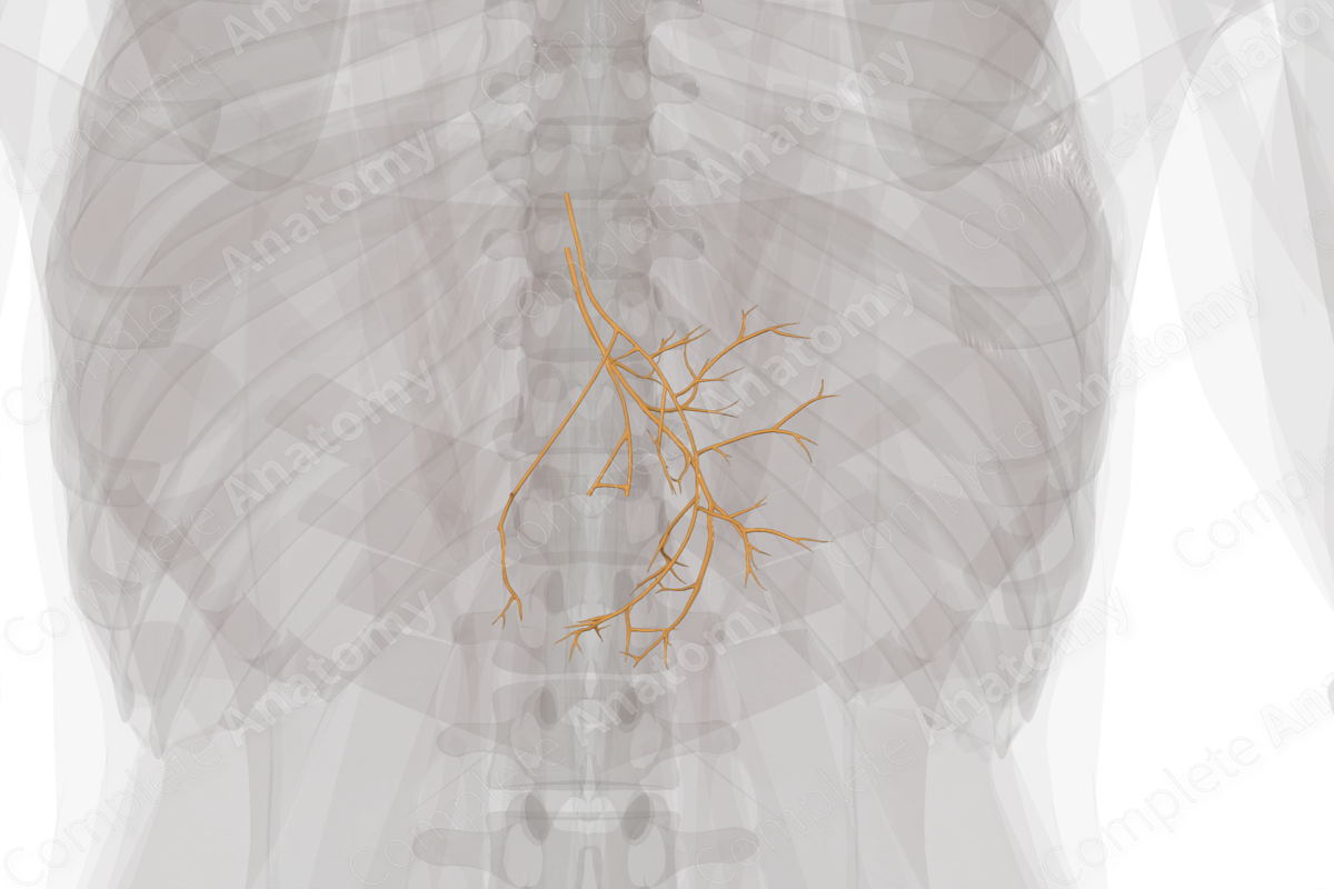
Description
The vagal part of the autonomic division refers to the parasympathetic fibers that contribute to the vagus nerve. These fibers are primarily associated with glandular secretion, slowing cardiac function, and increased contraction of smooth muscle in the digestive system. They have an extensive distribution.
In the pharynx and larynx, the vagal part of the autonomic division innervates small unnamed glands, the mucosa, and smooth muscle.
In the thorax, the vagal part of the autonomic division innervates smooth muscle and glandular tissue in the trachea, lungs, esophagus, and heart. Vagal input to the lungs results in bronchiole constriction and increased mucus production in the epithelium. Vagal fibers targeting the esophagus are responsible for the wave of smooth muscle contraction that propagates downward, propelling food to the stomach. Vagal fibers innervating the heart target the atrioventricular node, where they act to reduce the rate of cardiac contraction.
In the abdomen, the vagal part of the autonomic division is initially present as anterior and posterior vagal trunks, found on the anterior and posterior surfaces of the esophagus, just inferior to the esophageal hiatus. Branches of these trunks innervate the glands and smooth muscle of the digestive tract and accessory organs of the foregut and midgut. They also interface with the enteric nervous system found embedded in the walls of the foregut and midgut.
Visceral sensory fibers also travel along the vagus nerve, relaying sensory information from the viscera to the central nervous system. However, these fibers are not autonomic.
Related parts of the anatomy
Learn more about this topic from other Elsevier products


