Quick Facts
Sympathetic Contribution: Sacral splanchnic nerves.
Parasympathetic Contribution: Pelvic splanchnic nerves.
Course: Preganglionic axons travel along the inner surface of the sacrum into the inferior hypogastric plexus. From here they travel within the periarterial plexuses of the internal iliac, umbilical, superior, and inferior vesical arteries.
Sympathetic Supply: Inferior portion of the ureter, bladder wall (detrusor muscle), and internal urethral sphincter. In addition to the ductus deferens and seminal vesicle in males.
Parasympathetic Supply: Inferior portion of the ureter, bladder wall (detrusor muscle), and internal urethral sphincter, as well as the ductus deferens and seminal vesicle in males.


Contributing Nerves
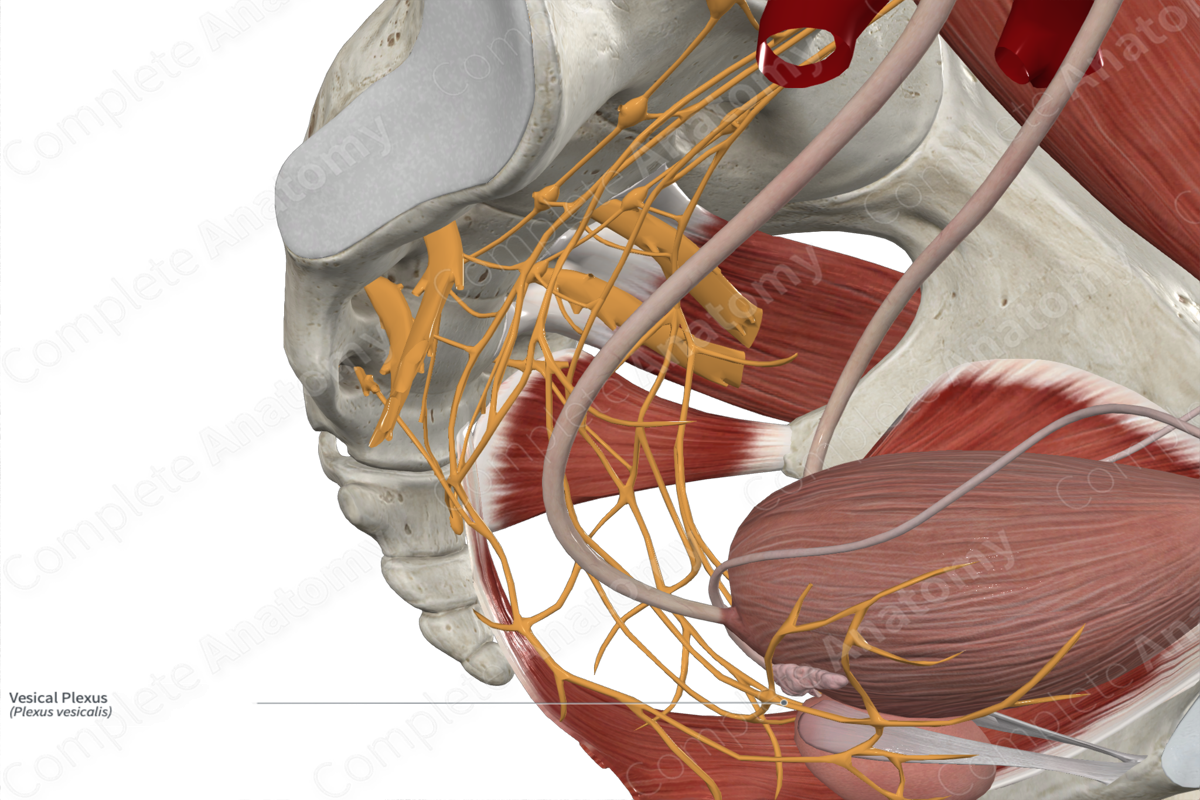
The sympathetic component of the vesical plexus gives rise to sympathetic sacral splanchnic nerves. The parasympathetic component gives rise to pelvic splanchnic nerves. Both become part of the inferior hypogastric plexus and most sympathetic fibers terminate on small ganglia scattered within the plexus. The vesical plexus is formed from a subset of these inferior hypogastric plexus nerves.
Course
The sympathetic component of this plexus arises from the eleventh thoracic to second lumbar (T11-L2) spinal cord segments. Preganglionic axons leave the cord and descend in the sympathetic trunk to the second to fourth sacral (S2-S4) levels. Here they peel off to become the sacral splanchnic nerves. These axons join parasympathetic preganglionic pelvic splanchnic nerves from S2-S4 spinal cord segments. Both sympathetic and parasympathetic nerves pass along the inner surface of the body of the sacrum, deep to the peritoneum, and become incorporated in the inferior hypogastric plexus.
Branches
Axons from the inferior hypogastric plexus ‘hitch a ride’ along the internal iliac artery. They pass from the periarterial plexus to their targets, or may contribute to those of the umbilical artery, the superior vesical branches of the umbilical artery, and the inferior vesical artery (males).
Supplied Structures
The vesical plexus provides autonomic innervation of the bladder (detrusor and internal urethral sphincter muscles), the pelvic portion of the ureter and, in males, the pelvic portion of the ductus deferens and seminal glands.
Visceral sensory fibers from the aforementioned organs are transmitted back towards the CNS through the vesical plexus.
List of Clinical Correlates
—Urinary continence
