Quick Facts
The arachnoid is a delicate membrane interposed between the dura mater and the pia mater, separated from the pia mater by the subarachnoid space (Dorland, 2011).
Structure and/or Key Features
There are three layers that make up the meninges, the dura, arachnoid, and pia mater. The meningeal layers can also be divided according to the stage of their development, including the leptomeninges (arachnoid and pia mater) and pachymeninx (dura mater).
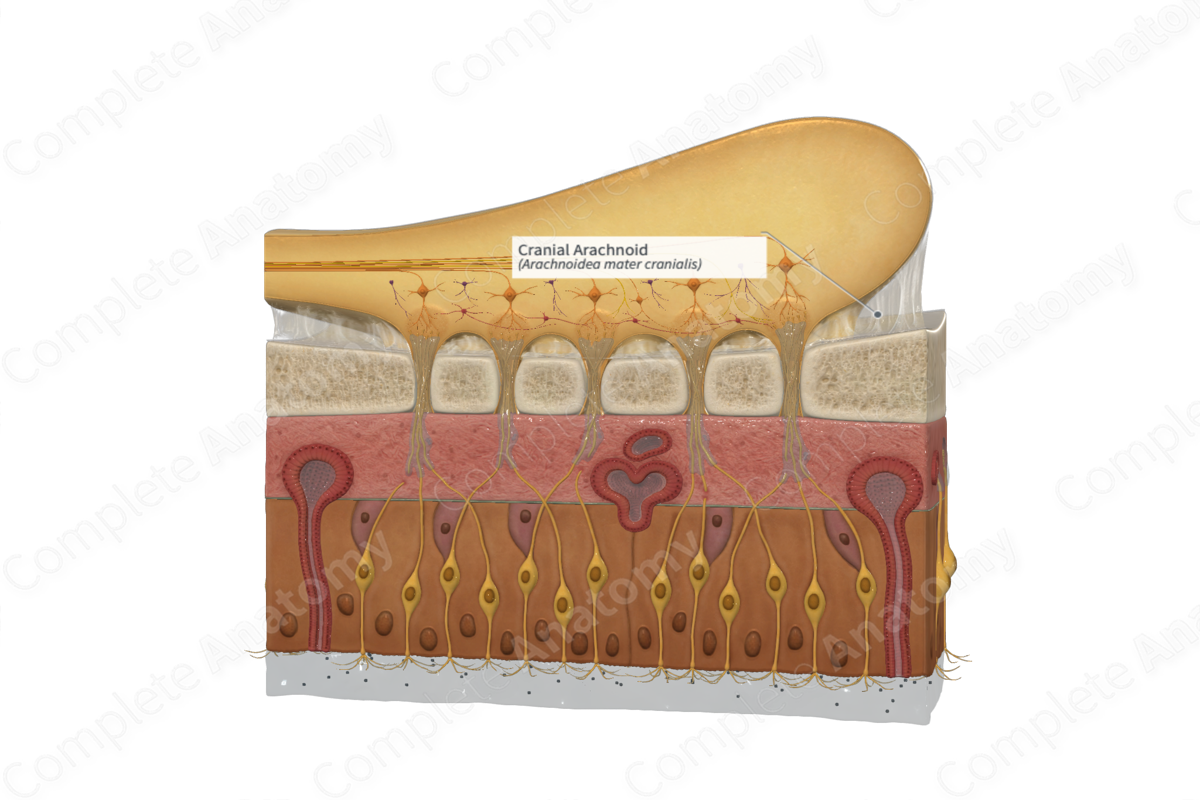
The arachnoid is a web-like structure made up of fibroblasts, collagen, and elastic fibers, and lies external to the pia mater and internal to the dura mater. It is separated from the pia by the subarachnoid space which contains cerebrospinal fluid.
The arachnoid is thicker than the pia mater, but thinner than the dura mater. Unlike the vascular, microscopic, and transparent pia mater, the arachnoid is an avascular layer that translucent, and thick enough to be handled with forceps.
The cranial aspect of arachnoid loosely encircles the surface of the brain, extending into the various sulci and fissures. The arachnoid is closely associated with the dura mater, but is not attached directly to it, due to the pressure exerted on it by the cerebrospinal fluid in the subarachnoid space. The arachnoid adheres to the adventitia of the internal carotid and vertebral arteries at the points where they make their entrance into the subarachnoid space.
The subarachnoid space is essentially a continuation of the lumen of the fourth ventricle and contains cerebrospinal fluid that is secreted by the choroid plexuses. It also contains a number of large blood vessels that travel across the surface of the brain. Cerebrospinal fluid usually flows within the subarachnoid space. Arachnoid villi and granulations, which are connected to the dural venous sinuses, reabsorbed cerebrospinal fluid, at intervals, back into the venous system.
Each branch of the olfactory nerve is enclosed by pia, arachnoid, and dura mater as it travels through the cribriform plate.
Anatomical Relations
The arachnoid lies external to the pia mater and internal to the dura mater. It is separated from the pia by the subarachnoid space which contains cerebrospinal fluid.
Function
The principal function of meningeal layers is to protect and provide support to the central nervous system.
The arachnoid plays a role in returning cerebrospinal fluid to the venous system, thus, maintaining correct intracranial pressure. Extensions of the arachnoid, called arachnoid villi, project beyond the dura mater into the dural venous sinuses.
Clinical Correlates
—Subarachnoid hemorrhage
—Subdural hemorrhage
—Bacterial meningitis
—Viral meningitis
—Meningioma
References
Dorland, W. (2011) Dorland's Illustrated Medical Dictionary. 32nd edn. Philadelphia, USA: Elsevier Saunders.
