Quick Facts
The lamina propria is the connective tissue coat of a mucous membrane just deep to the epithelium and basement membrane (Dorland, 2011).
Structure and/or Key Features
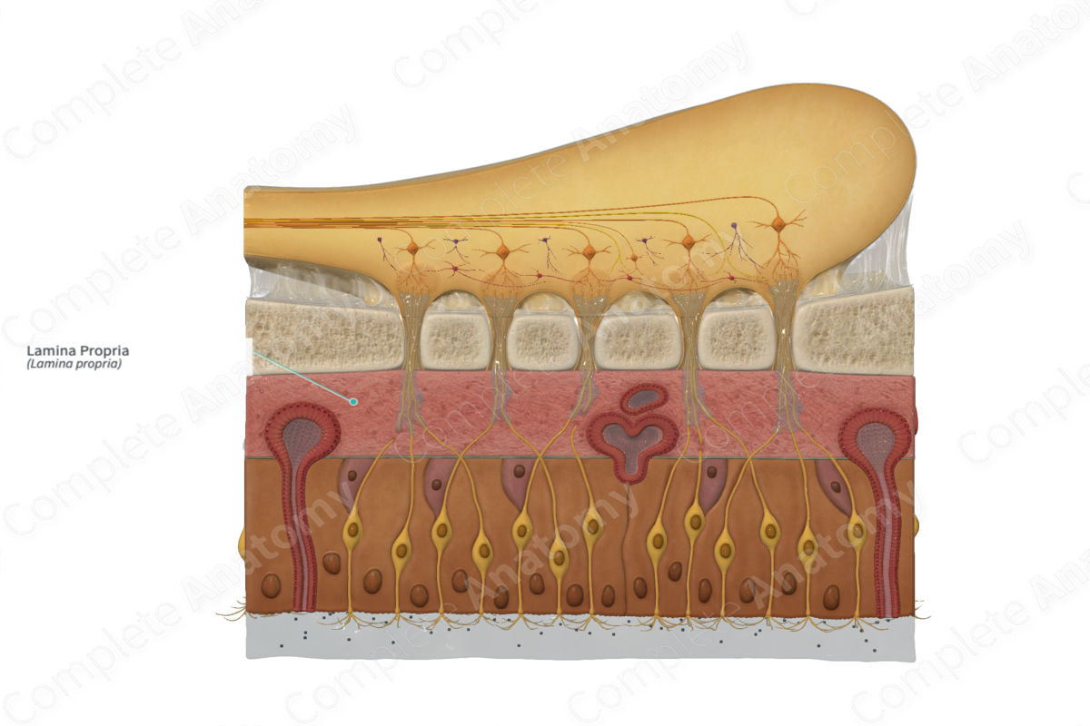
The olfactory mucosa is composed of the olfactory epithelium and the lamina propria.
The lamina propria is approximately two to three times thicker than the olfactory epithelium. It is situated deep to the olfactory epithelium, separated from it by a basement membrane.
The lamina propria is a highly vascularized layer made up of collagen, elastin, as well as a number of connective tissue elements including fibroblasts, macrophages, mast cells, and leukocytes. The lamina propria also contains lymphatic vessels, olfactory nerve fibers, and olfactory glands (Farbman et al., 1992; Krstic, 2013; Conn, 2008). A notable amount of olfactory ensheathing glia are situated within the lamina propria.
Anatomical Relations
The lamina propria is situated deep to the basement membrane of the olfactory epithelium, separating it from the cribriform plate of the ethmoid bone.
Function
The lamina propria contains blood and lymph vessels through which the olfactory organ is nourished and drained. It also contains immune cells such as macrophages, mast cells, and leukocytes. In addition to this, the lamina propria houses the olfactory glands, which are responsible for secreting the serous component of the mucus layer that covers the olfactory epithelium.
Clinical Correlates
—Anosmia
References
Conn, P. M. (2008) Neuroscience in Medicine. Humana Press.
Dorland, W. (2011) Dorland's Illustrated Medical Dictionary. 32nd edn. Philadelphia, USA: Elsevier Saunders.
Farbman, A. I., Bard, J. B. L., Barlow, P. W. and Kirk, D. L. (1992) Cell Biology of Olfaction. Developmental and Cell Biology Series: Cambridge University Press.
Krstic, R. V. (2013) Human Microscopic Anatomy: An Atlas for Students of Medicine and Biology. Springer Berlin Heidelberg.
