Quick Facts
The epineurium is the outermost layer of connective tissue of a peripheral nerve, surrounding the entire nerve and containing its supplying blood vessels and lymphatics (Dorland, 2011).
Structure/Morphology
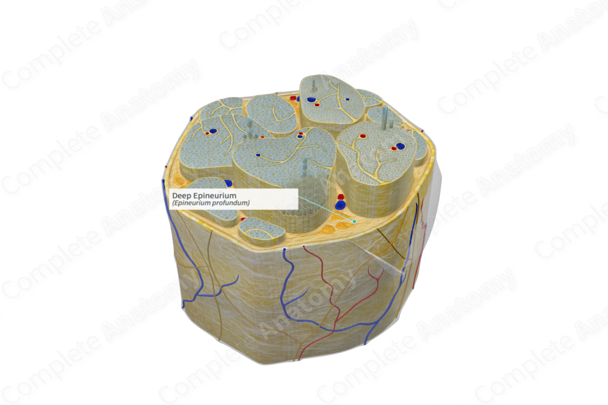
The deep (or internal) epineurium is located between the fascicles of the nerve trunk and is an inward extension of the external epineurium. It is composed of loose connective tissue, which includes fibroblasts, collagen (types I and III), and variable amounts of adipose tissue, which separate nerve fascicles from each other, within the nerve trunk (Standring, 2016). The deep epineurium also contains lymphatics and vasa nervorum, a network of fine blood vessels passing across the perineurium communicating with the endoneurium.
Key Features/Anatomical Relations
Condensed loose connective tissue arising from the mesoderm forms the epineurium. The epineurium is then divided into the superficial and deep epineurium. The deep epineurium separates individual fascicles from one another while the superficial epineurium surrounds the entire nerve.
Function
The deep epineurium, like the superficial epineurium, protects the internal nerve fibers. Connective tissue in its structure acts as a cushion, preventing compression or damage of the nerve. The deep epineurium also facilitates gliding between the fascicles (Barral and Croibier, 2007).
References
Barral, J. P. and Croibier, A. (2007) Manual Therapy for the Peripheral Nerves. Churchill Livingstone/Elsevier.
Dorland, W. (2011) Dorland's Illustrated Medical Dictionary. 32nd edn. Philadelphia, USA: Elsevier Saunders.
Standring, S. (2016) Gray's Anatomy: The Anatomical Basis of Clinical Practice. Gray's Anatomy Series: Elsevier Limited.
