Quick Facts
The epineurium is the outermost layer of connective tissue of a peripheral nerve, surrounding the entire nerve and containing its supplying blood vessels and lymphatics (Dorland, 2011).
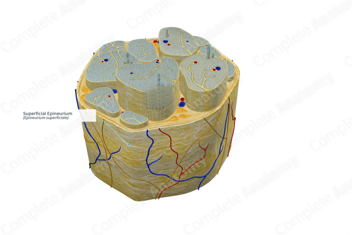
Structure/Morphology
The superficial (or external) epineurium is found encasing all fascicles of the nerve. It is about 60–100 nm thick. However thickness is related to the number of fasciculi present; the more fasciculi present the thicker the epineurium (Barral and Croibier, 2007). This structure contains fibroblasts, collagen fibers (types I and III), lymphocytes, and mast cells. The superficial epineurium also contains lymphatic vessels, vasa nervorum, and the nervi nervorum, which innervate the sheaths of nerve trunks (Standring, 2016).
Anatomical Relations
The epineurium is formed of loose connective tissue. It is divided into the superficial and deep epineurium. The superficial epineurium can be observed surrounding the nerve trunk, which contains multiple fascicles. It can be traced backwards to spinal meninges where spinal dura mater merges with the external epineurium just when the spinal nerve exits the intervertebral foramen. The deep epineurium refers to the layer of epineurium which penetrates within the nerve and separates the fascicles from one another (Snell, 2011).
Function
The superficial epineurium plays a role in protecting the internal nerve fiber it surrounds, cushioning it from damage e.g., during compression.
List of Clinical Correlates
—Pressure palsies in bedridden patients
References
Barral, J. P. and Croibier, A. (2007) Manual Therapy for the Peripheral Nerves. Churchill Livingstone/Elsevier.
Dorland, W. (2011) Dorland's Illustrated Medical Dictionary. 32nd edn. Philadelphia, USA: Elsevier Saunders.
Snell, R. S. (2011) Clinical Anatomy by Regions. Lippincott Williams & Wilkins.
Standring, S. (2016) Gray's Anatomy: The Anatomical Basis of Clinical Practice. Gray's Anatomy Series: Elsevier Limited.
