Quick Facts
The cone cell pedicle is the thick triangular or club-shaped ending of a retinal cone cell, which synapses with the bipolar and horizontal cells in the outer plexiform layer (Dorland, 2011).

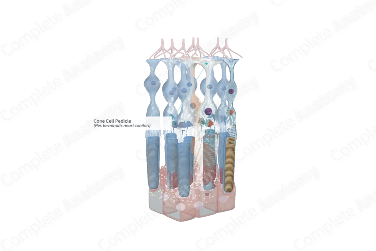
Structure and/or Key Features
Pedicles form the synaptic terminals of cone cells. They are distal expansions of the cone cell axon found at the junction of the outer nuclear and outer plexiform layers. They form the presynaptic part of connections with bipolar cells, horizontal cells, and other photoreceptor cells.
Measuring 8–10 µm in diameter, cone pedicles are described as large and conical, with the flat end facing the outer plexiform layer. Cone pedicles possess three distinct areas where neural synapses occur:
- invaginations in which are synaptic triads;
- flat surface connections with bipolar cells;
- lateral gap junction connections along the side of the pedicle with adjacent photoreceptor cells (Standring, 2016; Kolb, 1995).
Anatomical Relations
The cone cell pedicles are located in the outermost portion of the outer plexiform layer. Immediately lateral are neighboring cone pedicles, and, with increasing frequency from central to peripheral retina, the spherules of rod photoreceptors.
Function
The cone cell pedicles are responsible for making synaptic connections with bipolar cells, horizontal cells, and other photoreceptor cells. The photoreceptor cells release glutamate into the synapse.
Note that activation of photoreceptor cells in humans results in hyperpolarization, or inhibition, of that cell. Moreover, when photoreceptors are not activated, for example in a dark room, they remain in a depolarized state, thus constantly releasing the neurotransmitter glutamate. This release of glutamate at the synaptic terminal hyperpolarizes the connecting bipolar and horizontal cells; therefore, these cells are not activated, and the bipolar-ganglion synapse remains unexcited. When photoreceptors become activated by light, they hyperpolarize and the release of glutamate at the rod synaptic terminal ceases. The consequence of this is the depolarization of the connecting bipolar and horizontal cells and the transmission of the visual signal (Standring, 2016; Molday and Moritz, 2015).
References
Dorland, W. (2011) Dorland's Illustrated Medical Dictionary. 32nd edn. Philadelphia, USA: Elsevier Saunders.
Kolb, H. (1995) 'Photoreceptors', in Kolb, H., Fernandez, E. and Nelson, R. (eds.) Webvision: The Organization of the Retina and Visual System. Salt Lake City (UT): University of Utah Health Sciences Center. Copyright: (c) 2019 Webvision.
Molday, R. S. and Moritz, O. L. (2015) 'Photoreceptors at a glance', J Cell Sci, 128(22), pp. 4039-45.
Standring, S. (2016) Gray's Anatomy: The Anatomical Basis of Clinical Practice. Gray's Anatomy Series 41 edn.: Elsevier Limited.
