Quick Facts
The inner segment of a photoreceptor cell is the portion of a retinal rod or cone that is between the external limiting layer and the outer segment; this part contains the Golgi apparatus, endoplasmic reticulum, and numerous mitochondria (Dorland, 2011).

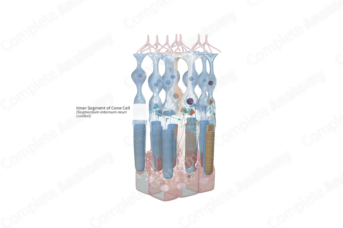
Structure and/or Key Features
Measuring approximately 5–6 µm in length, the inner segments of cone cells are generally much broader than that of rod cells. A general feature of inner segments of both rod and cone cells is that they may be divided into two regions, the myoid and the ellipsoid.
The myoid forms the proximal part of the inner segment, closest to the cell body, and houses the endoplasmic reticulum and Golgi apparatus. The ellipsoid forms the distal portion and contains densely packed mitochondria. Opsin proteins, such as those found in rhodopsin, are produced in the myoid but travel to the outer segment after production to be incorporated into newly forming membranous discs. Opsin proteins are integral components of the visual cycle. As new discs are added proximally, older discs move distally, toward the pigmented epithelium to be phagocytosed (Standring, 2016).
Anatomical Relations
The inner segments of cone cells form a retinal layer sublayer between the cone cell body (outer nuclear layer) and the outer segment layer.
Function
The inner segment of cone cells is highly specialized to supply the energy demand of these cells, and to support their requirements for protein synthesis. The inner segment of the photoreceptor cell is the primary site for these functions in photoreceptors.
References
Dorland, W. (2011) Dorland's Illustrated Medical Dictionary. 32nd edn. Philadelphia, USA: Elsevier Saunders.
Standring, S. (2016) Gray's Anatomy: The Anatomical Basis of Clinical Practice. Gray's Anatomy Series 41 edn.: Elsevier Limited.
