Quick Facts
The outer segments of rods and cones have flattened membranous discs which bear visual opsin.
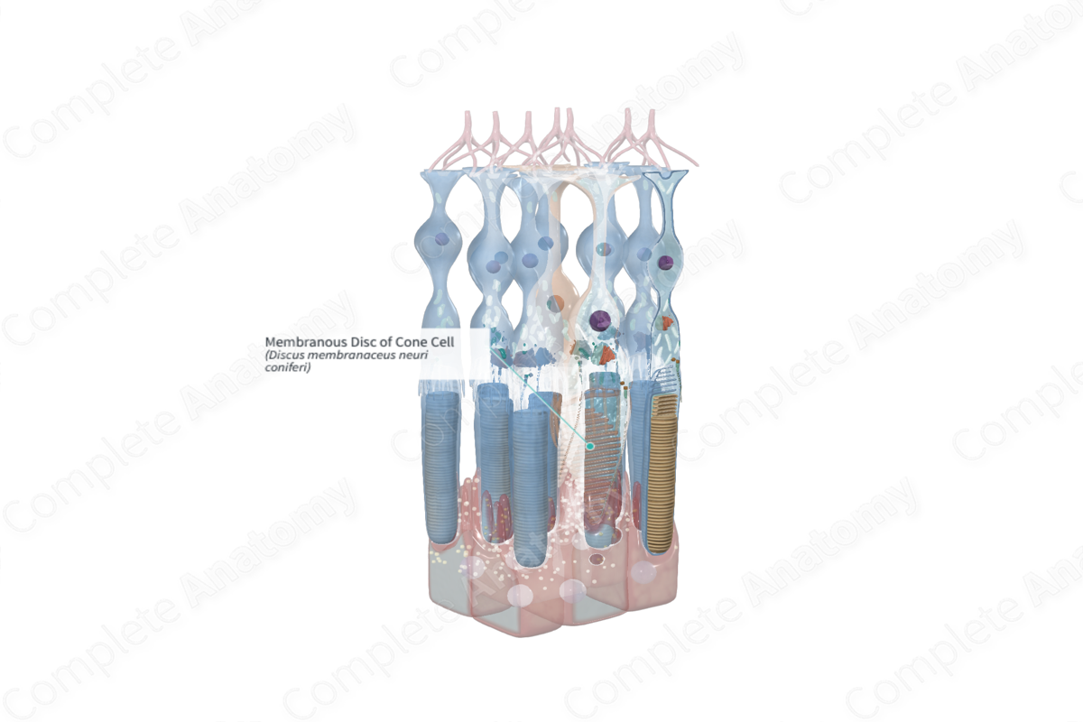
Structure and/or Key Features
The membranous discs of cone cells are present in the outer segment; however, they are less numerous than those in the rod cells and remain attached to the plasma membrane as infoldings at the base of the cell (Standring, 2016). There are additional reports that most, if not all, of the membranous discs in cone outer segments are continuous and remain attached to the plasma membrane, however, many of the connections appear to be very small. Therefore, it is assumed that intradiscal spaces are open to the interphotoreceptor matrix (Heckenlively, Arden and Bach, 2006).
A feature known as cone notches has also been described. These notches distinguish a site of transition between discs that are open to the interphotoreceptor matrix and those that are isolated (Heckenlively, Arden and Bach, 2006).
While rhodopsin is located on the outer segments of rod cells, a distinct feature of cone cells is the presence of proteins known as cone opsins or photopsins. There are three distinct photopsins, depending on the opsin protein that is present and bound to retinal (Standring, 2016). Each photopsin has a unique spectral sensitivity maximum and distribution.
Similar to rods, the membranous discs of cones are phagocytosed as new ones are manufactured. It should be noted that the incorporation of newly produced proteins in cone cells is more diffuse than that of rod cells.
Anatomical Relations
The membranous discs of rod cells are located in and extend the length of the outer segment of the rod photoreceptor cell.
Function
Despite their production in the inner segment of the cone cell, the spectrally distinct visual pigments are transported to the outer segments. The membranous discs are largely responsible for housing and anchoring the visual pigments. Following the absorption of light, a conformational change occurs in the opsin and retinal molecules resulting in the induction of a biochemical cascade which culminates in the production of an electrical signal representing visual information.
List of Clinical Correlates
- Rod-cone degeneration
- Rod-cone dystrophy
References
Heckenlively, J. R., Arden, G. B. and Bach, M. (2006) Principles and Practice of Clinical Electrophysiology of Vision. MIT Press.
Standring, S. (2016) Gray's Anatomy: The Anatomical Basis of Clinical Practice. Gray's Anatomy Series 41 edn.: Elsevier Limited.
