Quick Facts
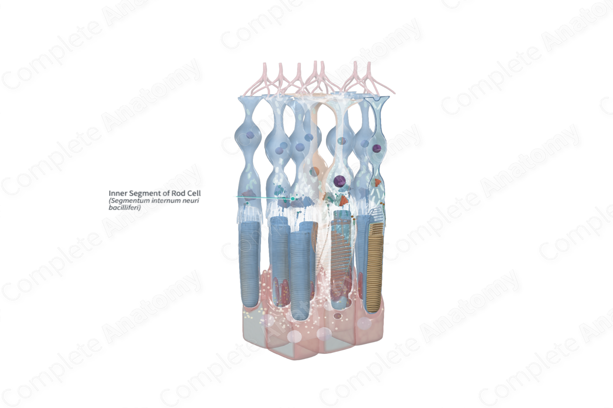
The inner segment of a photoreceptor cell is the portion of a retinal rod or cone that is between the external limiting layer and the outer segment; this part contains the Golgi apparatus, endoplasmic reticulum, and numerous mitochondria (Dorland, 2011).

Structure and/or Key Features
The inner segment is that portion of a photoreceptor is an organelle-rich cellular compartment that is external to the outer limiting membrane and internal to the connecting stalk. Measuring approximately 1.5 µm in length, the inner segment of rod cells is generally much narrower than the inner segment of cone cells, which measure approximately 5–6 µm. The inner segment of both rod and cone cells may be further divided into two distinct sections known as the myoid and the ellipsoid.
The myoid forms the basal part of the inner segment and houses the endoplasmic reticulum and Golgi apparatus. The ellipsoid forms the apical portion and contains densely packed mitochondria. Opsin proteins, such as those found in rhodopsin, are produced in the myoid but travel to the outer segment after production to be incorporated into the newly formed membranous discs. Opsin proteins are integral components of the visual cycle. As new discs are added, older discs get pushed toward the pigmented epithelium to be phagocytosed (Standring, 2016).
Anatomical Relations
The inner segments of the rod cells are largely located in the retinal layer of rods and cones (also known as the outer and inner segments of the retina).
Function
Photoreceptors are amongst the most metabolically active cells in the body. The inner segment of the photoreceptor cell is the primary site for protein synthesis and generating energy for the cell. Thus it is specialized with high density mitochondria to produce energy and Golgi apparatus, endoplasmic reticulum and ribosomes drive the protein synthesis.
References
Dorland, W. (2011) Dorland's Illustrated Medical Dictionary. 32nd edn. Philadelphia, USA: Elsevier Saunders.
Standring, S. (2016) Gray's Anatomy: The Anatomical Basis of Clinical Practice. Gray's Anatomy Series 41 edn.: Elsevier Limited.
