Membranous Disc of Rod Cell Quick Facts
The outer segments of rods and cones have flattened membranous discs which bear visual opsin.
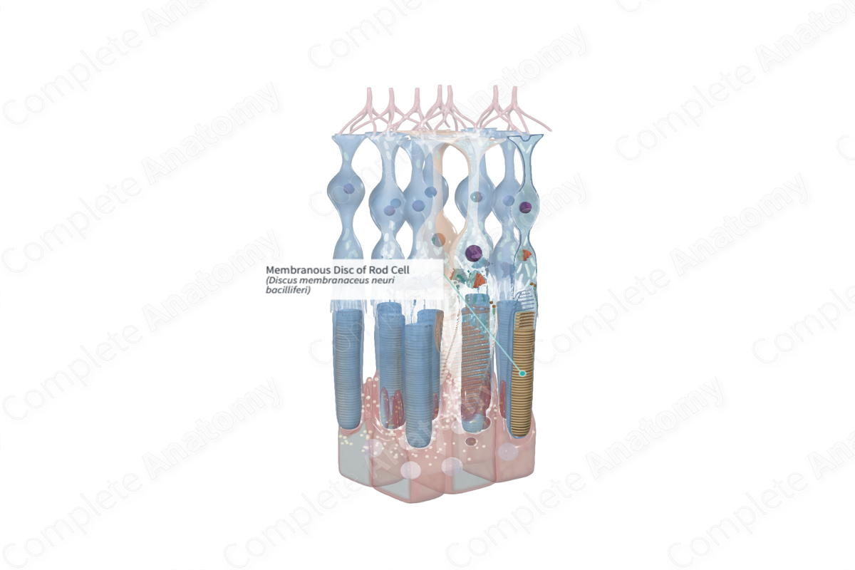
Membranous Disc of Rod Cell Structure and/or Key Features
The outer segments of rod cells are composed of approximately 1,000 stacked, flattened membranous discs, or lamellae. These discs originate as deep infoldings of the plasma membrane at the base of the outer segment. The bi-lipid membranous discs become detached from the surrounding plasma membrane following formation and are therefore free floating in the outer segment.
Embedded within the double membranes of the discs are light-sensitive visual pigment molecules, rhodopsin (Kolb, 1995; Standring, 2016).
There is constant turnover of membranous discs. From proximally, where they are formed at the base of the outer segments older discs are gradually displaced distally, up through the outer segment. At the very tip of the outer segment discs are pinched off, engulfed, and phagocytosed by the apical processes of the pigmented epithelium (Kolb, 1995).
Membranous Disc of Rod Cell Anatomical Relations
The multiple folded bilaminar membrane forming the discs of rod outer segments extend from the connecting stalk at their bases to the distal tip. The tips are surrounded by multiple microvillus processes of retinal pigment epithelium.
Membranous Disc of Rod Cell Function
Despite its production in the inner segment of the rod cell, rhodopsin subsequently migrates to the outer segments. The membranous discs of rod cells are largely responsible for housing and anchoring rhodopsin. Rhodopsin controls the absorption and processing of light under dim lighting conditions (Kolb, 1995). Most of the rhodopsin at the tips of rod outer segments are already photoconverted and so the no longer photosensitive pigment is sloughed off and engulfed, and phagocytized by the retinal pigment epithelium.
Membranous Disc of Rod Cell List of Clinical Correlates
- Rod-cone degeneration
- Rod-cone dystrophy
Membranous Disc of Rod Cell References
Kolb, H. (1995) 'Photoreceptors', in Kolb, H., Fernandez, E. and Nelson, R. (eds.) Webvision: The Organization of the Retina and Visual System. Salt Lake City (UT): University of Utah Health Sciences Center. Copyright: (c) 2019 Webvision.
Standring, S. (2016) Gray's Anatomy: The Anatomical Basis of Clinical Practice. Gray's Anatomy Series 41 edn.: Elsevier Limited.
