Quick Facts
The rod spherule is the pear-shaped ending of a retinal rod cell, which synapses with the bipolar and horizontal cells in the outer plexiform layer (Dorland, 2011).

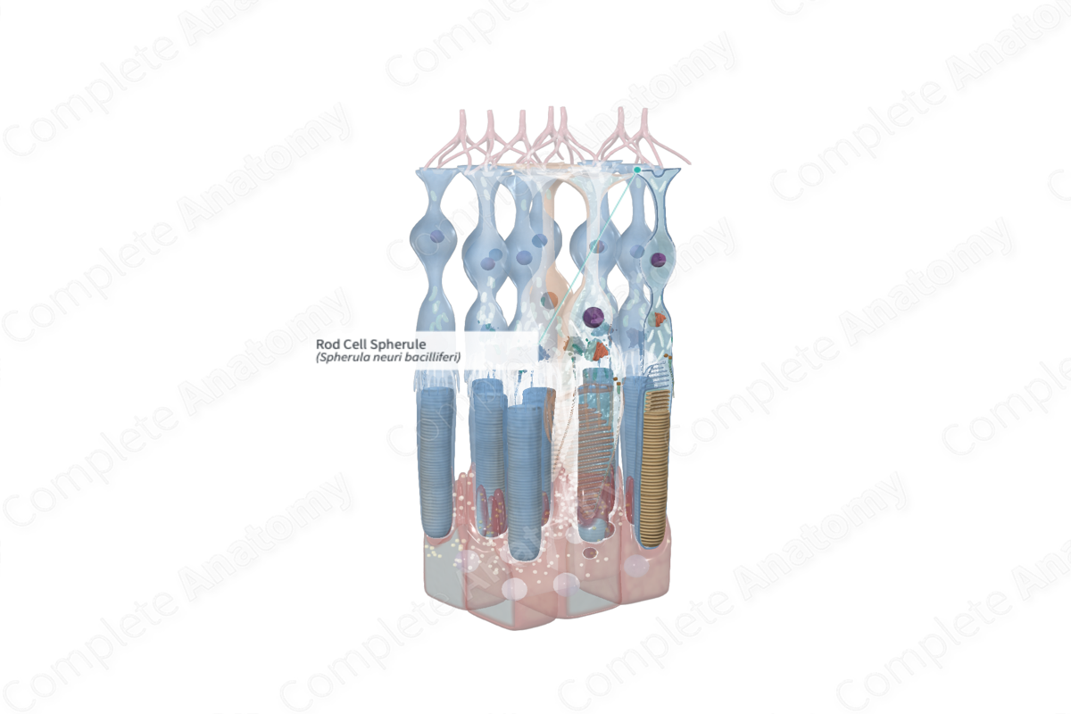
Structure and/or Key Features
The synaptic terminals of rod cells are oval to spherical and are called rod spherules. These expansions contain the presynaptic apparatus for connections with bipolar and horizontal cells. They also receive and make electrical synapses with neighboring cones (Csillag, 2007).
Each rod spherule exhibits one or more electron-dense structures, known as a synaptic ribbon. Synaptic ribbons are oriented generally perpendicular to the presynaptic membrane and are surrounded by many neurotransmitter containing vesicles (glutamate). The ribbon promotes the rapid migration of vesicles to the cell membrane and fusion with it to release its neurotransmitters (glutamate) into the synaptic cleft between the spherule and horizontal and bipolar cells (Standring, 2016; Kolb, 1995).
The basal membrane of rod spherules is invaginated by the postsynaptic processes of bipolar cells and horizontal cells in what is called and invaginated synapse. Rod spherules make connections with, on average, four bipolar cell processes. An individual horizontal cell contacts a spherule once, but one spherule may synapse with several horizontal cells. Typically there are a central bipolar cell elements flanked by horizontal cell elements (from separate cells) in an arrangement called a “synaptic triad.” (Boycott and Kolb, 1973).
Anatomical Relations
The rod cell spherules are located in the outer plexiform layer. Adjacent spherules are located at different depths within this layer. Spherules are surrounded by processes of bipolar and horizontal cells as well as the external processes of Müller glia that pass through the outer plexiform layer and outer nuclear layer before forming the outer limiting membrane of the retina.
Function
The rod cell spherules are responsible for making synaptic connections with bipolar and horizontal cells. The photoreceptor cells release glutamate into the synapse.
Note that activation of photoreceptor cells in humans results in hyperpolarization, or inhibition, of that cell. Moreover, when photoreceptors are not activated, for example in a dark room, they remain in a depolarized state, thus constantly releasing the neurotransmitter glutamate. This release of glutamate at the synaptic terminal hyperpolarizes the connecting bipolar and horizontal cells; therefore, these cells are not activated, and the bipolar-ganglion synapse remains unexcited. When photoreceptors become activated by light, they hyperpolarize and the release of glutamate at the rod synaptic terminal ceases. The consequence of this is the depolarization of the connecting bipolar and horizontal cells and the transmission of the visual signal (Standring, 2016; Molday and Moritz, 2015).
References
Boycott, B. B. and Kolb, H. (1973) 'The connections between bipolar cells and photoreceptors in the retina of the domestic cat', J Comp Neurol, 148(1), pp. 91-114.
Csillag, A. (2007) Atlas of the Sensory Organs: Functional and Clinical Anatomy. Humana Press.
Dorland, W. (2011) Dorland's Illustrated Medical Dictionary. 32nd edn. Philadelphia, USA: Elsevier Saunders.
Kolb, H. (1995) 'Photoreceptors', in Kolb, H., Fernandez, E. and Nelson, R. (eds.) Webvision: The Organization of the Retina and Visual System. Salt Lake City (UT): University of Utah Health Sciences Center. Copyright: (c) 2019 Webvision.
Molday, R. S. and Moritz, O. L. (2015) 'Photoreceptors at a glance', J Cell Sci, 128(22), pp. 4039-45.
Standring, S. (2016) Gray's Anatomy: The Anatomical Basis of Clinical Practice. Gray's Anatomy Series 41 edn.: Elsevier Limited.
