Quick Facts
A basement membrane is a thin sheet of amorphous extracellular material upon which the basal surfaces of epithelial cells rest; other cells associated with basement membranes are muscle cells, Schwann cells, and fat cells. The membrane is interposed between the cellular elements and the underlying connective tissue. It usually comprises two layers, the basal lamina and the reticular lamina, and is composed of Type IV collagen (which is unique to basement membranes), laminin, fibronectin, and heparan sulfate proteoglycans (Dorland, 2011).
Related parts of the anatomy
Structure and/or Key Feature(s)
Most epithelia attach to some form of underlying connective tissues by a basal lamina. The basal lamina is a thin sheet of extracellular proteins and fibrils. Additionally, carbohydrate-rich material is attached its lower face. If the accumulation is sufficient, the basal lamina is referred to as a basement membrane.
The basement membrane is synthesized by overlying epithelium and the underlying cells in connective tissue. Some epithelia do not have a basement membrane but most have a basal lamina (Ross and Pawlina, 2016; Ovalle, Nahirney and Netter, 2013).
Anatomical Relations
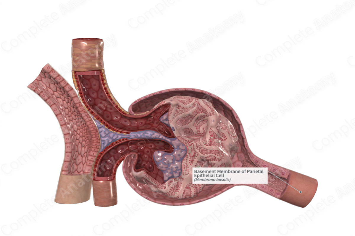
The parietal epithelial cells are attached to the underlying connective tissues by a very thin basement membrane.
Function
The basal lamina has a profound influence over the physiology of epithelium and a basement membrane adheres the epithelium to the underlying connective tissue being a supporting/strengthening component.
References
Dorland, W. (2011) Dorland's Illustrated Medical Dictionary. 32nd edn. Philadelphia, USA: Elsevier Saunders.
Ovalle, W. K., Nahirney, P. C. and Netter, F. H. (2013) Netter's Essential Histology. ClinicalKey 2012: Elsevier Saunders.
Ross, M. H. and Pawlina, W. (2006) Histology: A text and atlas. Lippincott Williams & Wilkins.
