Quick Facts
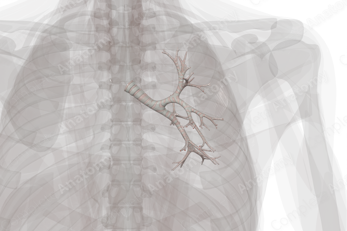
Location: Begin at the bifurcation of the trachea at the sternal angle (T4/T5) and enters the hilum of the left lung (T6).
Arterial Supply: Superior and inferior bronchial arteries.
Venous Drainage: Bronchial and pulmonary veins.
Innervation: Anterior and posterior pulmonary plexuses.
Lymphatic Drainage: Intrapulmonary lymph nodes.


Related parts of the anatomy
Structure
The bronchi arise from the bifurcation of the trachea into the right and left main bronchi at the level of the sternal angle (or transverse thoracic plane or angle of Louis). The left main bronchus bifurcates into superior and inferior lobar bronchi (or secondary bronchi) as it enters the hilum of the left lung, at the level of the sixth thoracic vertebra. These bronchi enter the superior and inferior lobes of the left lung, respectively.
Within the lobes, the lobar bronchi continue to divide into segmental (or tertiary) bronchi that supply each bronchopulmonary segment of the left lung.
Anatomical Relations
The main, lobar, and segmental (or primary, secondary, and tertiary) bronchi are accompanied by a pulmonary artery, which delivers deoxygenated blood to the lungs, and a pulmonary vein, which drains newly oxygenated blood from the lungs. Therefore, each lobe and each bronchopulmonary segment of the lung has its own bronchus, pulmonary artery, and adjacent intersegmental vein. This arrangement is important during surgery, since a single segment can be resected without affecting an adjacent segment.
Function
The primary function of the bronchi is to conduct air to the lungs so that gas exchange and oxygenation of blood can occur. As the bronchi are constantly exposed to inhaled allergens, particles, and microbes, it is necessary to clear these threats to maintain homeostasis. The mucosa of the bronchi and bronchioles play the vital role in protecting the lungs from these environmental insults, thus, maintaining homeostasis (Ganesan, Comstock and Sajjan, 2013).
Arterial Supply
The bronchial arteries supply the oxygenated blood and nutrients to the bronchi. A paired group of left bronchial branches arise directly from the descending thoracic aorta, the superior and inferior bronchial branches.
Venous Drainage
The bronchial veins receive blood from the bronchi, hilar structures, such as lymph nodes, and visceral pleura adjacent to the hilum. The left bronchial vein arises from the posterior surface of the left bronchi. It travels posteriorly to its terminations at the hemiazygos vein, or sometimes the superior intercostal vein. Additionally, some blood from the bronchial arteries is drained by the pulmonary veins. Please see our micro-anatomy model of the bronchial tree for more details.
Innervation
The bronchi receives autonomic innervation from the anterior and posterior pulmonary plexuses. The nerves of the pulmonary plexuses originate from the thoracic branches of the upper sympathetic trunk (sympathetic contribution) and the vagus nerve (parasympathetic contribution). For both sympathetic and parasympathetic fibers, axons may travel directly to the pulmonary plexus, or may travel through the cardiac plexus first. The autonomic innervation of the trachea controls airway function, such as tone of smooth muscle, mucus secretion, and vascular permeability and blood flow.
Lymphatic Drainage
The intrapulmonary lymph nodes are located along the bronchi. These nodes drain the deep bronchopulmonary plexus located within the bronchopulmonary segments of the lungs. The intrapulmonary lymph nodes send efferents to the bronchopulmonary lymph nodes at the hilum of the lungs.
References
Ganesan, S., Comstock, A. T. and Sajjan, U. S. (2013) 'Barrier function of airway tract epithelium', Tissue Barriers., 1(4), pp. e24997.
Learn more about this topic from other Elsevier products
Trachea and bronchi histology: Video, Causes, & Meaning

Trachea and bronchi histology: Symptoms, Causes, Videos & Quizzes | Learn Fast for Better Retention!
