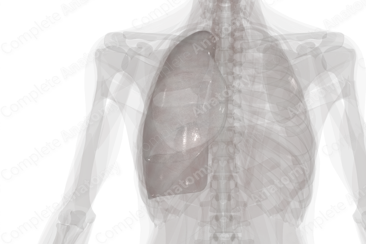
Pleura (Right Lung) Quick Facts
Location: Surrounds right lung.
Arterial Supply: Internal thoracic, intercostal, and bronchial arteries, branches of the subclavian artery.
Venous Drainage: Azygos system, pulmonary and bronchial veins.
Innervation: Pulmonary plexus, anterior rami of thoracic nerves (or intercostal nerves), and branches of phrenic nerve.
Lymphatic Drainage: Superior diaphragmatic, parasternal, intercostal, and mediastinal lymph nodes, superficial pulmonary lymphatic plexus.


Pleura (Right Lung) Structure
The pleura consists of two layers, an inner visceral and an outer parietal layer. Both layers form a closed sac called the pleural cavity, which contains serous fluid. Due to the position of the heart, the left pleural cavity is smaller than the right.
The parietal pleura lines the thoracic wall, diaphragm, and mediastinum, while the inner visceral pleura closely invests the surface of the lungs and sits within the fissures of the lung. In cadaveric dissection, it is not possible to separate the visceral pleura from the lung tissue. The visceral and parietal pleura are continuous at the hilum where they form the pulmonary ligament.
Pleura (Right Lung) Anatomical Relations
The parietal layer of pleura is a continuous structure; however, it is divided into regions according to its anatomical relations. The costal (or costovertebral) pleura lines the ribs and vertebrae; the diaphragmatic pleura lines the diaphragm; the cervical pleural lines the apex of the lungs; the mediastinal pleura lines the medial aspects of the lungs.
The relations of the mediastinal pleura are different in the right and left lungs indicative of the asymmetries of the mediastinal structures. The most profound difference between left and right pleura are due to the positioning of the heart on the left. Specifically, the sternal border of the pleura of the left lung is displaced laterally by the left ventricle of the heart.
Pleura (Right Lung) Function
Within the pleural cavity, a thin layer of fluid is produced by the serous epithelial membrane lining the visceral and parietal pleurae. In addition, the visceral pleura invests all surfaces of the lung and gives the lung a shiny appearance. This smooth surface and fluid layer allow friction-free movement during respiration. Additionally, the fluid in the pleural cavity provides surface tension, akin to water between two pieces of glass, that keeps the lung surface in contact with the thoracic wall (Moore, Dalley and Agur, 2013). This permits the parietal pleura to adhere to the inner thoracic wall, the diaphragm, and the mediastinum, and thus, keep the lungs inflated.
Pleura (Right Lung) Arterial Supply
The parietal pleural receives its arterial supplied according to its position within the thoracic cavity. The costal pleura receives vascular supply from the arteries that supply the thoracic wall, including the intercostal and internal thoracic arteries. The mediastinal pleura is supplied via bronchial vessels. The diaphragmatic pleura receives its supply from the vessels supplying the diaphragm, while the cervical pleura is supplied by branches of the subclavian artery.
The visceral pleura is supplied, along with the lungs, via the bronchial arteries. At the hilum of each lung, the bronchial arteries branch around the primary bronchus and pleural branches arise from here. They supply the mediastinal visceral pleura, the interlobar surfaces, the apex, and a portion of the diaphragmatic visceral pleura.
Pleura (Right Lung) Venous Drainage
Venous drainage of the parietal pleura occurs via thoracic wall veins which drain into the azygos system. Venous drainage of the visceral pleura occurs primarily via pulmonary and bronchial veins.
Pleura (Right Lung) Innervation
The parietal pleura is innervated according to its position within the thoracic cavity. The costal and diaphragmatic parietal pleurae are innervated by the intercostal nerves (or anterior rami of the thoracic nerves) and pain is associated with the corresponding portion of the thoracic or abdominal wall. The mediastinal and central diaphragmatic parietal pleura are innervated by the phrenic nerves and pain may be referred to the tip of the shoulder i.e., dermatome C3-C4. The visceral pleura receives afferents from the pulmonary plexus that course with the bronchial vessels (Standring, 2016).
Pleura (Right Lung) Lymphatic Drainage
Lymph from the parietal pleura drains into the superior diaphragmatic, parasternal, intercostal, and mediastinal lymph nodes. Lymph from visceral pleura is drained via the superficial pulmonary lymphatic plexus.
Pleura (Right Lung) List of Clinical Correlates
- Pneumothorax
- Hydrothorax
- Hemothorax
- Chylothorax
- Pleurectomy
- Pleurisy
Pleura (Right Lung) References
Moore, K. L., Dalley, A. F. and Agur, A. M. R. (2013) Clinically Oriented Anatomy. Clinically Oriented Anatomy 7th edn.: Wolters Kluwer Health/Lippincott Williams & Wilkins.Standring, S. (2016) Gray's Anatomy: The Anatomical Basis of Clinical Practice. Gray's Anatomy Series 41 edn.: Elsevier Limited.
Learn more about this topic from other Elsevier products
Pleurae

Pleura covers the lung and interior of the thorax and the pleural cavity is the thin fluid-filled space between the two pulmonary pleurae (known as visceral and parietal) of each lung.
