Quick Facts
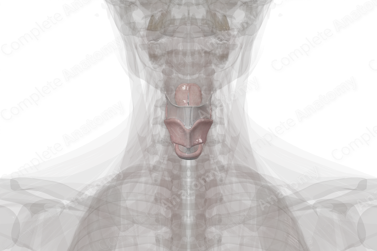
Location: In the neck at the level of the third to sixth cervical vertebrae.
Arterial Supply: Laryngeal branches of superior and inferior thyroid arteries.
Venous Drainage: Superior and inferior laryngeal veins.
Innervation: Recurrent laryngeal nerve (CN X) and superior laryngeal nerve (CN X).
Lymphatic Drainage: Prelaryngeal, thyroid, paratracheal lymph nodes.


Structure
The skeletal framework of the larynx is composed of nine cartilages (three paired cartilages and three unpaired cartilages). The cartilages are connected to each other by intrinsic membranes and to surrounding structures by extrinsic membranes.
Several extrinsic and intrinsic muscles permit movement of the larynx as a whole or as individual parts of the larynx (i.e., epiglottis and vocal folds).
The cartilages, membranes, and muscles are coated by a mucosa, the outer layer that is seen clinically during a laryngoscopic examination.
Anatomical Relations
The larynx is a mobile structure, and, at rest, it is typically located at the level of C3—C6 vertebrae in males. This varies slightly in children and in females due to the larger size of the post-pubertal male larynx. As a consequence of high testosterone levels, the male larynx increases in size until age 40 years (Standring, 2016).
The larynx is relatively superficial as it is only in part covered by some infrahyoid (strap) muscles and the thyroid gland.
Function
The larynx has many functions including a conduit for air, a sphincter to close off the airways, and is involved in the process of phonation and deglutition.
Arterial Supply
The vascular supply to the larynx arises from the laryngeal branches of the superior and inferior thyroid arteries. The superior laryngeal artery travels with the internal branch of the superior laryngeal nerve as it pierces the thyroid hyoid membrane. A branch of the superior laryngeal artery, the cricothyroid artery, supplies the cricothyroid muscle. The inferior laryngeal artery travels with the inferior laryngeal nerve (i.e. the terminal portion of the recurrent laryngeal nerve) to supply the mucosa and muscles of the inferior larynx.
Venous Drainage
The larynx is drained by the superior and inferior laryngeal veins, which unit with the superior and inferior thyroid veins, respectively.
Innervation
Innervation of the larynx is completed by branches of the vagus nerve (CN X).
Motor innervation to the larynx arises primarily from the recurrent laryngeal nerve (the anterior terminal branch), except for the cricothyroid muscle. The cricothyroid muscle is innervated by the external laryngeal nerve (a branch of the superior laryngeal nerve).
The recurrent laryngeal nerve also has a sensory component (the posterior terminal branch) which supplies the inferior region of the larynx, below the vocal folds. The region superior to the vocal folds receives sensory input from superior laryngeal nerve. However, considerable overlap between these nerves is likely.
Lymphatic Drainage
Lymphatic drainage of the region superior and inferior to the vocal folds occurs via lymph vessels traveling with the superior and inferior thyroid arteries, respectively. These lymph vessels drain in the superior and inferior deep cervical lymph nodes.
Additionally, lymph from the inferior aspect of the larynx may also drain via paratracheal nodes before draining into the inferior deep cervical lymph nodes.
References
Standring, S. (2016) Gray's Anatomy: The Anatomical Basis of Clinical Practice. Gray's Anatomy Series 41 edn.: Elsevier Limited.
Learn more about this topic from other Elsevier products

Anatomy of the larynx and trachea: Video, Causes, & Meaning

Anatomy of the larynx and trachea: Symptoms, Causes, Videos & Quizzes | Learn Fast for Better Retention!
