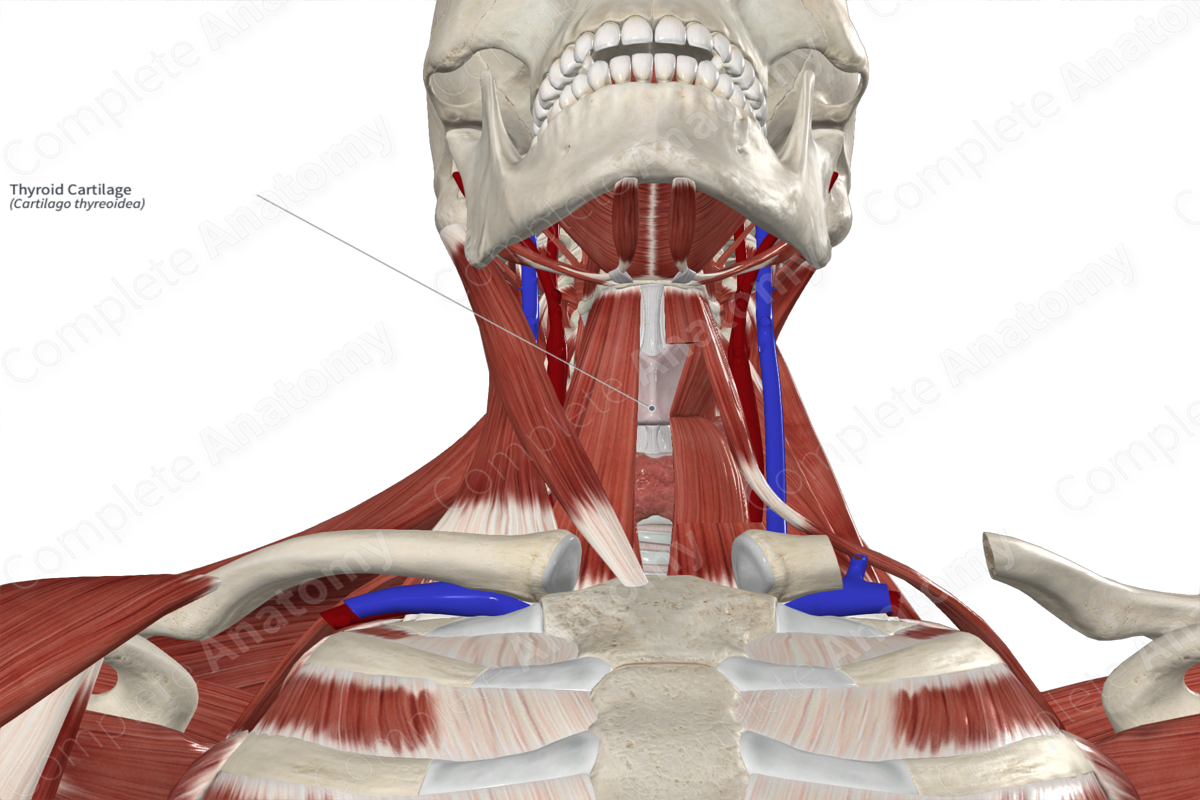
Structure
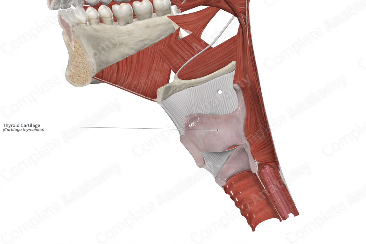
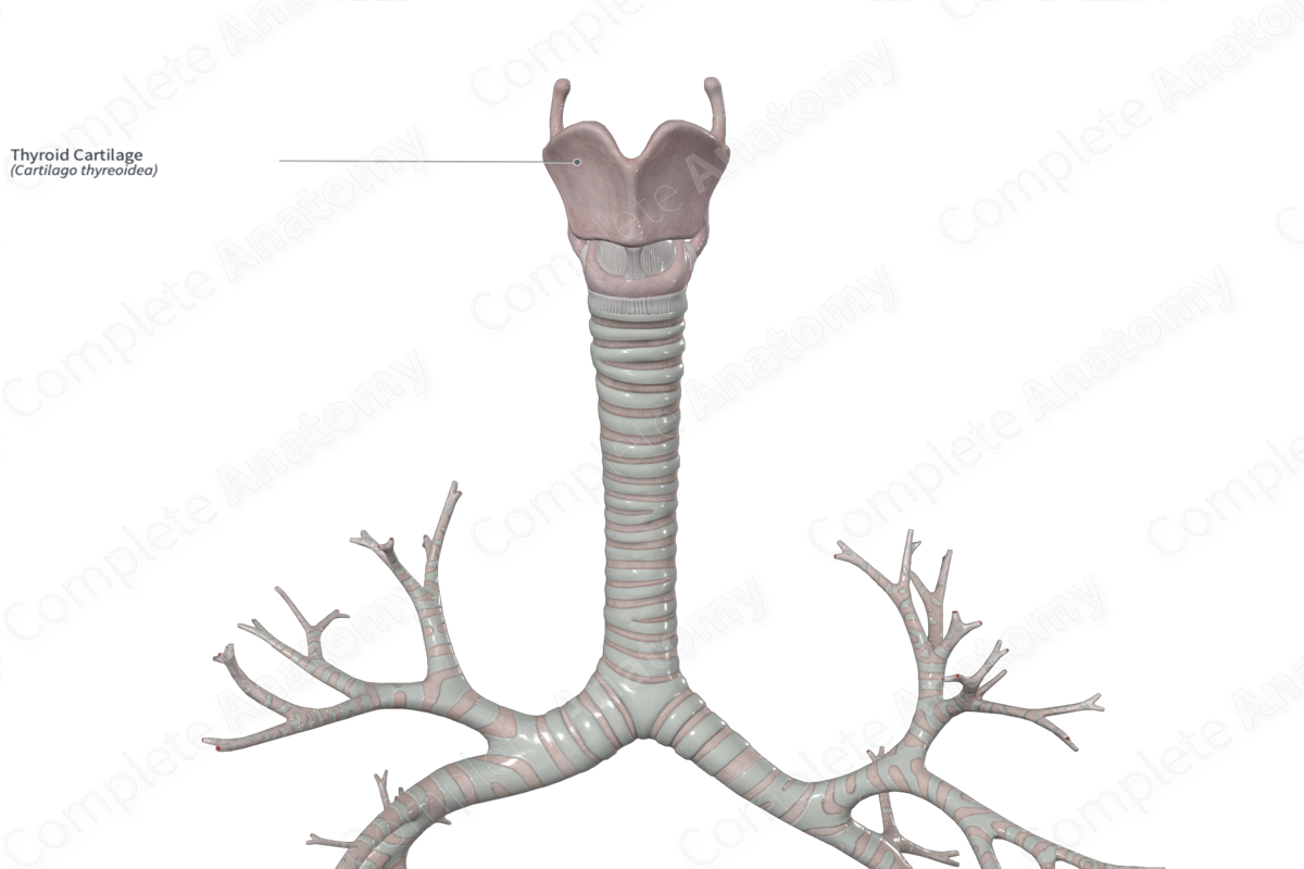
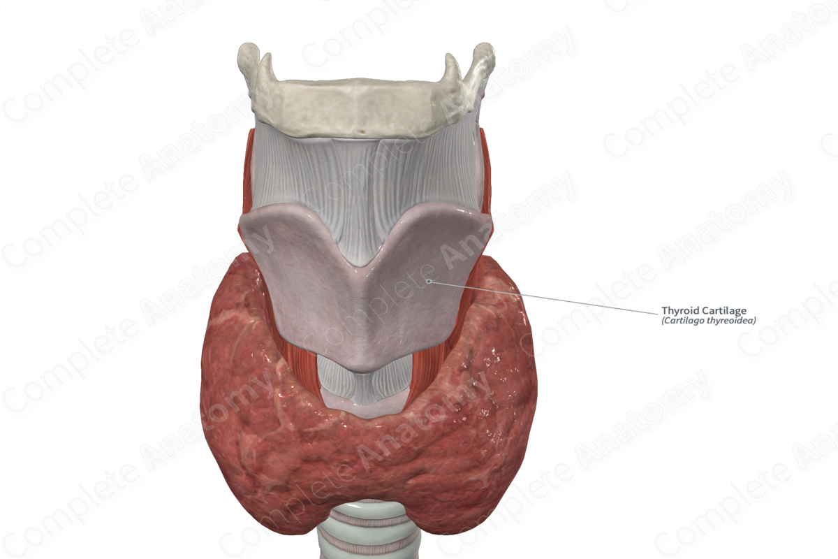
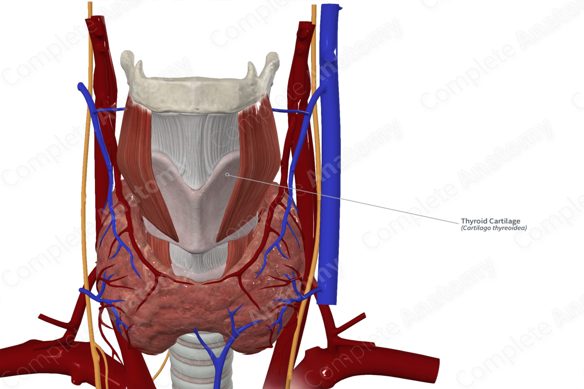
The thyroid cartilage is composed of hyaline cartilage. It is a large C-shaped cartilage that is deficient posteriorly. It has two anteriorly placed laminae. The inferior two thirds of these broad laminae fuse medially. At the superior-most point of this fusion is the laryngeal prominence, which is colloquially termed the “Adam’s apple.” The angle at which the laminae fuse is approximately 90 degrees in males and 120 degrees in females. Thus, the angle in males is associated the more pronounced laryngeal prominence, longer vocal cords, and a deeper pitched voice (Standring, 2016).
Above the laryngeal prominence is the superior thyroid notch.
The oblique line can be seen as a ridge on the external aspect of the anterior surface. This is the site of attachment for two infrahyoid muscles, the sternothyroid and the thyrohyoid muscles, as well as the thyropharyngeal portion of the inferior constrictor muscle of the pharynx.
Posteriorly, the two laminae have a superior and an inferior projection, named the superior and inferior horns.
Key Features & Anatomical Relations
The superior portion of the thyroid cartilage is connected to the hyoid bone via the thyrohyoid ligament while, the shorter and thicker inferior horns articulate with the cricoid cartilage below.
The thyroid cartilage is attached to the epiglottis via the thyroepiglottic ligament. Additionally, it attaches both the vestibular and vocal ligaments and thus provides the necessary skeletal framework required for the formation of the true and false vocal folds. It also serves as the site of attachment for some of the intrinsic laryngeal muscles.
References
Standring, S. (2016) Gray's Anatomy: The Anatomical Basis of Clinical Practice. Gray's Anatomy Series 41 edn.: Elsevier Limited.
Learn more about this topic from other Elsevier products
Thyroid Cartilage

The thyroid cartilage is the largest cartilage of the larynx and is formed by the union of broad, paired lamina.

