Quick Facts
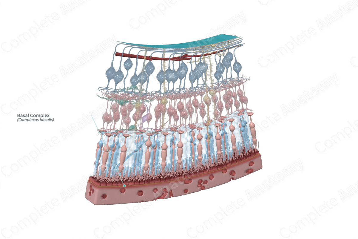
The basal complex is the transparent inner layer of the choroid, which is in contact with the pigmented layer of the retina (Dorland, 2011).
Related parts of the anatomy
Structure and/or Key Features
The basal complex, otherwise known as the lamina vitreal or Bruch’s membrane, is a barrier lying between the choroidocapillary and the retinal pigment epithelium. It is 2–4 µm thick and is composed of a central elastic layer flanked by inner and outer fibrous layers of collagen and basement membrane (basal lamina).
Anatomical Relations
The basal complex is a thin layer that, at its outer surface abuts the overlying choroidocapillary lamina of the choroid. At its inner surface, it separates this lamina from the retinal pigment epithelium. A potential space, the remnant of the lumen of the embryonic optic vesicle, is present between these two and is a potential site of retinal detachment.
Function
The basal complex allows for the passage of nutrients, gases, and metabolites between the dense capillary bed of the choroidocapillary and the avascular outer layers of the retina. With age, it can be the site where deposits become embedded. This can impair the passage of substances between the choroid and retina and contribute to retinal photoreceptor degeneration.
List of Clinical Correlates
- Retinal detachment
- Drusen
- Age-related macular degeneration
References
Dorland, W. (2011) Dorland's Illustrated Medical Dictionary. 32nd edn. Philadelphia, USA: Elsevier Saunders.
