Quick Facts
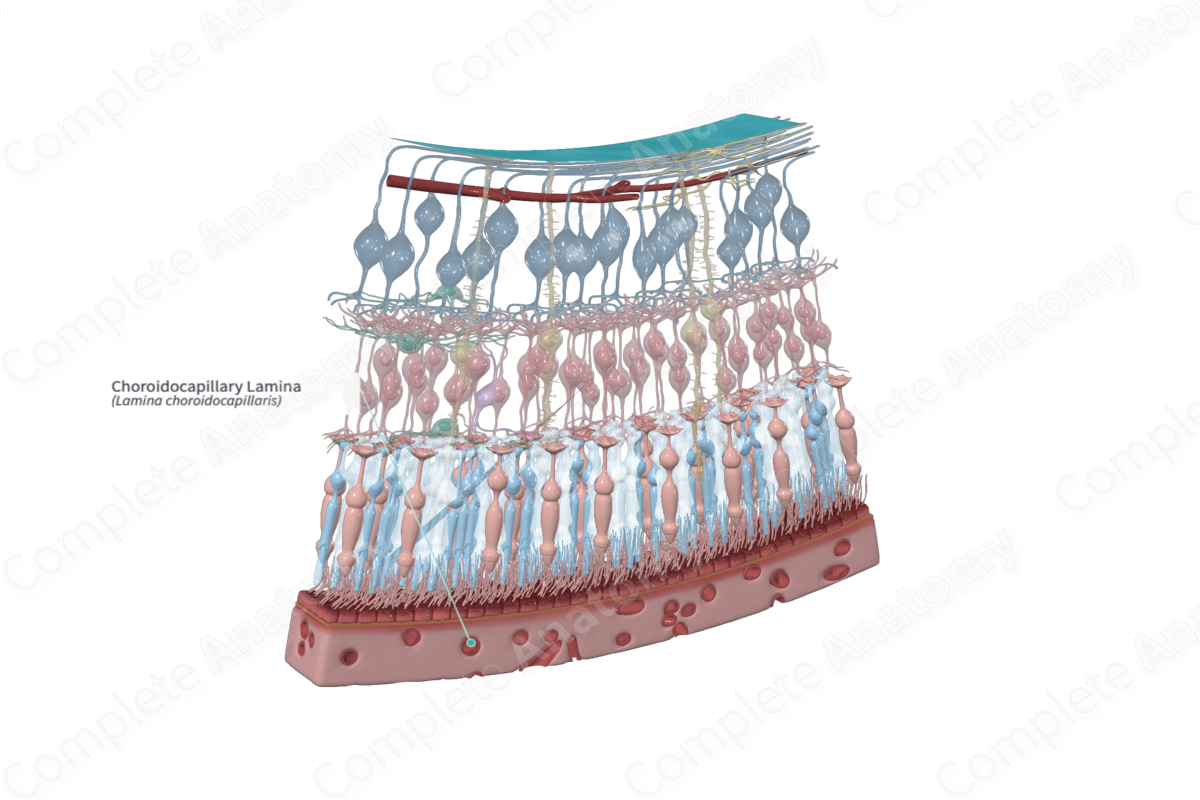
The choroidocapillary lamina is the inner layer of the choroid, composed of a single-layered network of small capillaries (Dorland, 2011).
Related parts of the anatomy
Structure and/or Key Features
The choroidocapillary lamina forms the innermost layer of the choroid. It is composed of densely packed, large fenestrated capillaries that are freely anastomosing. This layer is so highly vascularized that the light reflected from the back of the eye in flash photography is colored an intense red.
Anatomical Relations
The choroidocapillary lamina is situated between the basal complex (lamina vitrea or Bruch’s membrane) and the vascular layer in the choroid. The basal complex separates the choroidocapillary lamina from the retina.
Function
The vascular beds of the choroidocapillary lamina are vital for supplying the adjacent retina. The outer segments of the photoreceptors, the outer nuclear layer and the outer plexiform layer (avascular layers), in that order, are immediately adjacent to the choroidocapillary layer and depend entirely on diffusion from it for oxygen and nutrients.
List of Clinical Correlates
- Retinal detachment
- Age-related macular degeneration
References
Dorland, W. (2011) Dorland's Illustrated Medical Dictionary. 32nd edn. Philadelphia, USA: Elsevier Saunders.
