Quick Facts
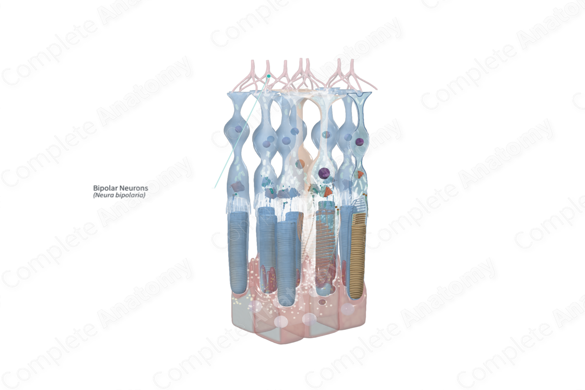
A bipolar cell is a nerve cell with two processes. Bipolar neurons located in the inner nuclear layer of the retina are the second neurons in the sensory pathway, making synaptic contacts with photoreceptors and retinal ganglion cells (Dorland, 2011).

Structure and/or Key Features
Appropriately named, bipolar neurons are recognizable by two distinct projections, one axon and one dendrite, attached to a cell body. Known as the second-order neurons in the visual pathway, bipolar neurons are responsible for relaying information from photoreceptors cells to ganglion cells (Remington and Goodwin, 2011).
In mammals, bipolar neurons are designated as cone bipolar cells or rod bipolar cells, depending on which photoreceptor cell they receive input from. There are approximately 10 distinct forms of cone bipolar cells but only one form of rod bipolar cell (Remington and Goodwin, 2011; Strettoi et al., 2010).
According to size and connectivity, cone bipolar cells may be largely categorized as midget cone, blue cone, or diffuse cone bipolar cells. A subtype of midget bipolar cells, known as flat midget bipolar cells, synapses on the base of the cone pedicle. Most other midget bipolar cells make contact via direct invagination of the cone pedicle. In the peripheral retina, a midget bipolar cell may contact 2–3 cone cells. However, in the central retina, each midget bipolar cell synapses with just one cone photoreceptor cell, forming a direct channel from photoreceptor to ganglion cells, facilitating high spatial resolution (Standring, 2016).
Another distinction between bipolar cells may be made from their response to light stimulation. There are ON bipolar cells and OFF bipolar cell differing in their response to the light-induced hyperpolarization of photoreceptor cells and the consequent cessation of glutamate release. Bipolar cells that depolarize following the activation and hyperpolarization of photoreceptor cells are classified as ON cells. On the other hand, bipolar cells that hyperpolarize following the light-induced hyperpolarization of photoreceptor cells are known as OFF cells (Standring, 2016).
Rod bipolar cells all share a single morphology. One rod bipolar cell will contact 40–45 rods in the peripheral retina and 30–35 rods in the central retina. Interestingly, bipolar rod cells do not directly contact ganglion cells, but instead make axodendritic or axoaxonic synapses with amacrine cells which proceed to excite ON retinal ganglion cells and inhibit OFF retinal ganglion cells by dendrodendritic connections. Thus on electron micrographs, one can see an arrangement called a serial synapse whereby a bipolar cell terminal is presynaptic to an amacrine terminal, which itself is presynaptic to a retinal ganglion cell terminal (Standring, 2016; Bloomfield and Dacheux, 2001).
Anatomical Relations
Bipolar neurons are radially orientated. Their cell bodies are located in the inner nuclear layer. Within this layer they tend to be in the outer and middle portion, adjacent and vitreal to the horizontal cells. They are more sparse in the inner portion, which is mostly composed of Müller glia. The orientation of bipolar cells is perpendicular to the retinal layers. One process serves as a dendrite extending into the outer plexiform layer and is postsynaptic to photoreceptor cells, horizontal cells, and interplexiform cells. From the opposite side of the cell body, an axonal process extends into the inner plexiform layer where it is presynaptic to amacrine cells processes (Standring, 2016).
Function
Bipolar cells are largely responsible for the direct and indirect transmission of visual signals from photoreceptor cells to ganglion cells. Specifically, the dendrites of bipolar cells receive input from photoreceptor cells, horizontal cells, and interplexiform cells and synapse directly onto ganglion cells or indirectly via amacrine cells (Remington and Goodwin, 2011).
Following the light-induced hyperpolarization of photoreceptor cells, the release of glutamate at the synaptic clefts is inhibited. ON bipolar cells will depolarize in response to the inhibition of glutamate, while OFF bipolar cells will hyperpolarize. As aforementioned, rod bipolar cells do not directly make contact with ganglion cells, but instead synapse with amacrine cells which excite surrounding ON bipolar cone cells and inhibit OFF bipolar cone cells. This results in an overriding of the cone pathway and facilitates the transmission of scotopic rod signals to ganglion cells in ambient light conditions (Standring, 2016; Bloomfield and Dacheux, 2001).
List of Clinical Correlates
- Paraneoplastic retinopathy
- Congenital stationary night blindness
References
Bloomfield, S. A. and Dacheux, R. F. (2001) 'Rod vision: pathways and processing in the mammalian retina', Prog Retin Eye Res, 20(3), pp. 351-84.
Dorland, W. (2011) Dorland's Illustrated Medical Dictionary. 32nd edn. Philadelphia, USA: Elsevier Saunders.
Remington, L. A. and Goodwin, D. (2011) Clinical Anatomy of the Visual System E-Book. Elsevier Health Sciences.
Standring, S. (2016) Gray's Anatomy: The Anatomical Basis of Clinical Practice. Gray's Anatomy Series 41 edn.: Elsevier Limited.
Strettoi, E., Novelli, E., Mazzoni, F., Barone, I. and Damiani, D. (2010) 'Complexity of retinal cone bipolar cells', Prog Retin Eye Res, 29(4), pp. 272-83.
