Quick Facts
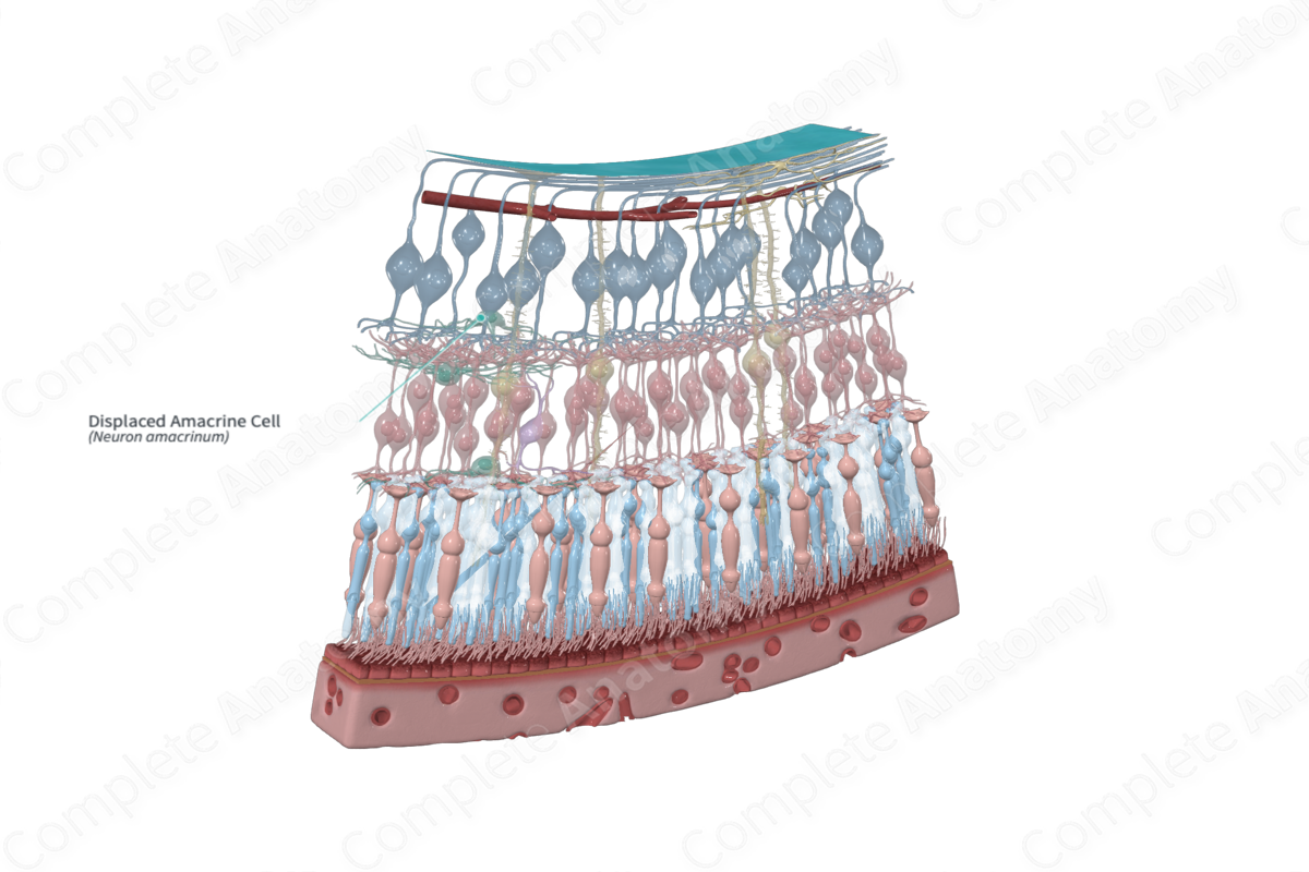
An amacrine cell is a type of multipolar neuron, located in the inner nuclear layer of the retina, that lacks an axon (Dorland, 2011). The cell bodies of displaced amacrine cells are located in the ganglionic layer of the retina.
Structure and/or Key Features
Amacrine cells are described as interconnecting neurons, or interneurons. Interneurons are a specific type of neuron that act as intermediaries, responsible for influencing the transfer of information between two projection neurons. Amacrine cells are specifically classified as inhibitory interneurons (Standring, 2016). Most amacrine cell bodies are located in the inner nuclear layer, but a population is “displaced” such that their cell bodies are found in the retinal ganglion cell layer.
Amacrine cells are recognizable by their large cell bodies and lobulated nuclei. They do not have a typical axon, with cell extensions acting both pre- and post-synaptically. Some possess a single long process that ends with extensive branching. The long process electrotonically isolates those processes emerging from the cell body from those at the distal end of this long process, thus allowing these cells to act proximal and distal to the amacrine cell body. Amacrine cell processes extend into the inner plexiform layer where it synapses with bipolar cells, ganglion cells, and other amacrine cells (Standring, 2016; Remington and Goodwin, 2011).
It is currently estimated that there are approximately 30–40 different amacrine cell types. Amacrine cells may be categorized depending on their process morphology and stratification (stratified or diffuse). They may be further classified depending on the spread or reach of their processes (narrow-field, small-field, medium-field, and large-field). Additionally, they may be classified by the type of inhibitory neurotransmitter they release. The diversity of amacrine cell types is reflected in the complexity and scale of their signaling role within the retina (Remington and Goodwin, 2011; Balasubramanian and Gan, 2014).
Anatomical Relations
The cell bodies of displaced amacrine cells are located in the retinal ganglion cell layer intermixed with the ganglion cells. They can be distinguished from them by the lack of an axon that extends in the nerve fiber layer, to the optic disc and into the optic nerve. Thus, displaced amacrine cells cannot be backfilled by a retrograde tracer injected into the visual centers of the brain, nor do they exhibit Wallerian degeneration due to severing of the optic nerve. The processes of displaced amacrine cells extend externally into the inner plexiform layer (Standring, 2016).
Function
Amacrine cells are diverse in morphology and function not all of which are understood. Some are involved in the detection of directional motion, others regulate the eyes’ ability to adapt to light, and others still play a role in establishing the body’s circadian rhythm (Balasubramanian and Gan, 2014).
Like horizontal cells, amacrine cells are known to be inhibitory. They receive excitatory input from bipolar cells and subsequently integrate and regulate the information that reaches ganglion cells via feedback synapses onto bipolar terminals or via feedforward synapses onto ganglion cell dendrites. Amacrine cells may also communicate, via lateral inhibitory synapses, with other amacrine cells (Diamond, 2017).
Amacrine cells play an important role in shaping the visual signaling transmitted through the retina. For example, inhibitory feedback signals from amacrine cells to bipolar cells sharpen the timing of bipolar cell responses and additionally refines their center-surround receptive fields. Feedforward signals, on the other hand, ensure there is spatial and temporal detail in the receptive field (Diamond, 2017).
References
Balasubramanian, R. and Gan, L. (2014) 'Development of Retinal Amacrine Cells and Their Dendritic Stratification', Curr Ophthalmol Rep, 2(3), pp. 100-106.
Diamond, J. S. (2017) 'Inhibitory Interneurons in the Retina: Types, Circuitry, and Function', Annu Rev Vis Sci, 3, pp. 1-24.
Dorland, W. (2011) Dorland's Illustrated Medical Dictionary. 32nd edn. Philadelphia, USA: Elsevier Saunders.
Remington, L. A. and Goodwin, D. (2011) Clinical Anatomy of the Visual System E-Book. Elsevier Health Sciences.
Standring, S. (2016) Gray's Anatomy: The Anatomical Basis of Clinical Practice. Gray's Anatomy Series 41 edn.: Elsevier Limited.
