Quick Facts
A horizontal cell is a retinal neuron; there are two types, and their functions are unclear. Each cell has a multipolar soma in the internal nuclear layer and one long neurite and several short ones. All the neurites serve as both axons and dendrites, extending along and ramifying within the internal nuclear layer. The long neurites synapse in the outer plexiform layer with both pedicles and spherules; the short neurites synapse either with pedicles or with spherules (Dorland, 2011).
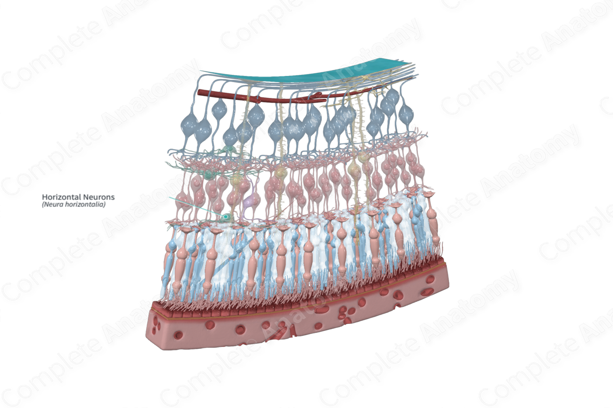
Structure and/or Key Features
Horizontal neurons are described as interconnecting neurons that transfer information in a lateral or horizontal direction. Interconnecting neurons, or interneurons, are specific types that act as an intermediary role in the transfer of information between two projection neurons. Horizontal cells are specifically classified as inhibitory interneurons (Standring, 2016).
Horizontal cells have a cell body located at the external margin of the inner nuclear layer, a single long axon, and several shorter dendrites with branching terminals. They synapse with bipolar and photoreceptor cells. Additionally they are connected to neighboring horizontal cells by gap junctions forming an extensive, interconnected network (Remington and Goodwin, 2011).
There are three morphologically distinct types of horizontal neurons in the retina, commonly called H1, H2, and H3 horizontal cells. The dendrites of H1 and H3 horizontal neurons form synaptic connections with cone cells, while their axons connect with rod cells. On the other hand, both axons and dendrites of H2 horizontal neurons form synaptic connections with cone cells only (Standring, 2016; Kolb, 1995c).
Anatomical Relations
The cell bodies of horizontal neurons are located along the external margin of the inner nuclear layer of the retina. Their long processes extend out along the junction between the outer plexiform and inner nuclear layers, running parallel to the retinal surface. They subsequently enter the outer plexiform layer and terminate there.
Function
Horizontal neurons are largely responsible for helping the eyes to adjust under both bright and dim light. They additionally have a role in contrast enhancement and distinguishing opposing colors (Twig, Levy and Perlman, 2003).
Horizontal neurons are described as inhibitory interneurons. When photoreceptor cells transduce light energy into a neural signal that is transmitted to bipolar and ganglion cells, horizontal cells provide feedback and feedforward inhibition to photoreceptor and bipolar cells, respectively. In the retina, this is known as lateral inhibition. As such, horizontal neurons help to integrate and regulate signals from photoreceptor cells (Diamond, 2017). It should be noted that considering photoreceptors have an intrinsic response whereby light-evoked “activation” of the cell results in hyperpolarization, the inhibitory feedback provided by horizontal neurons results in the depolarization of photoreceptor cells.
H1 horizontal neurons have dendrites that synapse with approximately 7–18 cones, while their axons terminate at the rod spherule. In this sense, H1 neurons facilitate communication and integration of signals between rod and cone cells. In this way, they play a role in the shift from scotopic (night) to photopic (day) vision and vice versa. Similar to H1 neurons, H3 neurons have a large dendritic tree that contacts many cones. Interestingly, H3 neurons seem to avoid blue cones; therefore, they are likely selective for red and green cones. The termination of the H3 axon remains unclear but are likely to contact both rod and cone cells. H2 neurons have processes, both dendrites and axons, that simply connect cones and are believed
References
Diamond, J. S. (2017) 'Inhibitory Interneurons in the Retina: Types, Circuitry, and Function', Annu Rev Vis Sci, 3, pp. 1-24.
Dorland, W. (2011) Dorland's Illustrated Medical Dictionary. 32nd edn. Philadelphia, USA: Elsevier Saunders.
Kolb, H. (1995c) 'Photoreceptors', in Kolb, H., Fernandez, E. and Nelson, R. (eds.) Webvision: The Organization of the Retina and Visual System. Salt Lake City (UT): University of Utah Health Sciences Center.
Remington, L. A. and Goodwin, D. (2011) Clinical Anatomy of the Visual System E-Book. Elsevier Health Sciences.
Standring, S. (2016) Gray's Anatomy: The Anatomical Basis of Clinical Practice. Gray's Anatomy Series 41 edn.: Elsevier Limited.
Twig, G., Levy, H. and Perlman, I. (2003) 'Color opponency in horizontal cells of the vertebrate retina', Prog Retin Eye Res, 22(1), pp. 31-68.
