Quick Facts
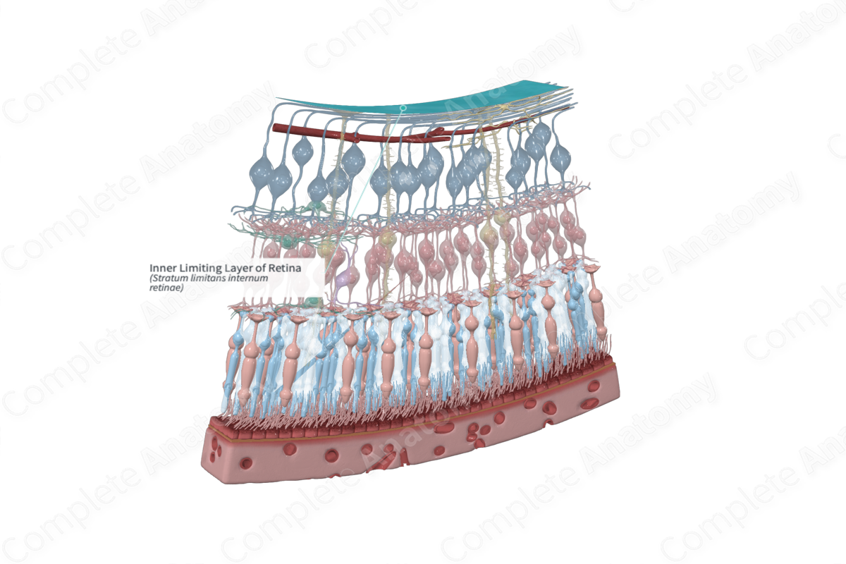
The inner limiting layer of the retina is the basal lamina of the Müller fibers in the retina; it is the layer of the neural retina located just internal to the layer of nerve fibers and separates the inner, conical ends of the cells from the vitreous body (Dorland, 2011).
Structure and/or Key Features
The inner limiting layer, or membrane, is described as the innermost boundary of the retina and occurs at the interface between the optic nerve fiber layer of the retina and the vitreous body. The outer, retinal surface of the membrane is uneven, which can be attributed to the inner processes of radial glial (Müller) cells that terminate in a foot plate known as the “endfoot.” Endfeet of Müller cells tile together like pieces of a jigsaw puzzle forming a continuous sheet. The inner, vitreal surface of the limiting membrane has a smooth appearance that is covered by a membrane, with some vitreous fibers apparent in the periphery (Remington and Goodwin, 2011).
At the margins of the optic disc, the inner limiting membrane undergoes modification, where the processes of Müller cells are replaced by those belonging to astrocytes (Remington and Goodwin, 2011). Unlike the outer limiting membrane, the inner limiting membrane is a true basement membrane and is estimated to 0.5-2 μm thick in the posterior retina (Standring, 2016).
Anatomical Relations
The inner limiting membrane lies superjacent to the layer of retinal ganglion cell axons and is the most vitreously positioned layer of the retina. Anteriorly, it is continuous with the inner limiting membrane of the ciliary body (Remington and Goodwin, 2011).
Function
The inner limiting membrane is responsible for creating a boundary between the retina and the vitreous body. Furthermore, it acts as a diffusion barrier between the two structures (Kolb, 1995c).
List of Clinical Correlates
- Macular holes
References
Dorland, W. (2011) Dorland's Illustrated Medical Dictionary. 32nd edn. Philadelphia, USA: Elsevier Saunders.
Kolb, H. (1995c) 'Photoreceptors', in Kolb, H., Fernandez, E. and Nelson, R. (eds.) Webvision: The Organization of the Retina and Visual System. Salt Lake City (UT): University of Utah Health Sciences Center.
Remington, L. A. and Goodwin, D. (2011) Clinical Anatomy of the Visual System E-Book. Elsevier Health Sciences.
Standring, S. (2016) Gray's Anatomy: The Anatomical Basis of Clinical Practice. Gray's Anatomy Series 41 edn.: Elsevier Limited.
