Quick Facts
The pigmented layer of retina is a layer of pigmented epithelium that is the outer of the two parts of the optic part of the retina, extending from the entrance of the optic nerve to the pupillary margin of the iris. It is important in the turnover of rods and cones and functions as a blood-retina barrier to maintain the ionic environment of the retina (Dorland, 2011).
Structure and/or Key Features
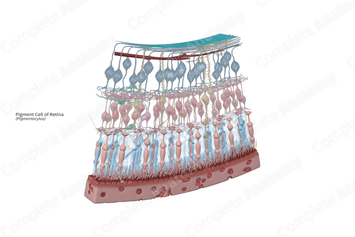
The outermost layer of the retina, the pigmented layer, is a composed of pigment cells arranged as a continuous, single row of cuboidal epithelial cells. There are approximately 4–6 million pigment cells in the retina. These cells tend to have a flat appearance in radial section and a hexagonal or pentagonal appearance in surface view (Standring, 2016).
Pigment cells contain multiple organelles called melanosomes, cytoplasmic granules that facilitate the synthesis, storage, and transportation of melanin. Melanin is a common light-absorbing pigment in biology. In retinal pigment cells, melanosomes are retained intracellularly; thus melanosomes and melanin facilitate the absorption of light that has passed through the neural retina to this outer epithelial layer minimizing reflection back to the neural retina, as this would degrade the resolution of an image (Wasmeier et al., 2008).
Pigment cells possess actin-rich apical processes, or microvilli, that are approximately 5–7 µm long. These microvilli interdigitate between the outer segments of the photoreceptors internal to them (Wasmeier et al., 2008). The basal surface of pigment cells is highly invaginated, which increases the surface area of the pigmented layer for the absorption of nutrients. The lateral membranes of the pigment cells are firmly connected by tight junctions, which collectively form the outer blood-retinal barrier (Boulton and Dayhaw-Barker, 2001).
The shape, size, and appearance of pigment cells varies depending on their location within the retina. Cells in the macula measure approximately 12 µm long and 14 µm in diameter. Moving away from the macula, cells become wider and flatter, with some cells near the ora serrata measuring approximately 60 µm in diameter (Boulton and Dayhaw-Barker, 2001).
Anatomical Relations
The pigment cells extend from the periphery of the optic disc to the ora serrata, where it is continuous with the outer ciliary epithelium. The pigment cells lie between the choroid’s basal complex (Bruch’s membrane) and the layer of rods and cones.
Embryologically, the retina is derived from the two layers of the invaginated optic vesicle. The outer layer develops into the layer of pigment cells which lies between and separates the neural retina and the choroid. The inner layer of the optic vesicle forms the other nine layers of the retina collectively known as the neural layer (Standring, 2016). Due to differences in the embryological origins of the pigmented layer and the nine layers beneath it, the border between these two areas is not supported by junctional complexes. Therefore, the neural layers and the pigmented layer of the retina can easily become detached, commonly seen in the instance of trauma and disease; this is known as retinal detachment.
Function
The function of the pigment cells is the maintenance and protection of the adjacent photoreceptor cells. They are also thought to play an important role in the integrity of the choroidal capillaries (Boulton and Dayhaw-Barker, 2001).
The pigment cells form and act as the outer blood-retinal barrier. As such, it regulates the transepithelial transport of ions, fluid, and metabolites that reach the photoreceptors via specialized transport proteins and lateral plasma membrane barrier junctions. Further, glucose is passively transported, and amino acids are actively absorbed across this epithelial layer, while waste products are removed (Boulton and Dayhaw-Barker, 2001).
Another essential role carried out by the pigment cells is in the transport and storage of retinoids. Retinoids are described as analogs of vitamin A, which are fundamental in the maintenance of the visual cycle. The pigment cells are additionally responsible for the ingestion and degradation of the spent outer segments of photoreceptors via phagocytosis (Boulton and Dayhaw-Barker, 2001).
The pigmented layer plays a critical role in the protection of photoreceptors against light and free radicals. Specifically, the intensity and wavelength of light that reaches the photoreceptors are regulated by macular pigments and melanin which respectively function to filter out reactive blue light and act as a neutral density filter to reduce light passing the pigmented layer. Enzymatic and non-enzymatic antioxidants are also contained within the pigmented layer which neutralizes reactive oxygen species, preventing damage to cellular molecules (Boulton and Dayhaw-Barker, 2001).
Pigment cells also have a phagocytic function. The microvilli that surround the photoreceptor outer segments will “pinch off,” engulf and phagocytose those at the distal tips that have exhausted the photopigment by transduction. This process occurs in a diurnal rhythm producing a turnover for about 7–10% of photoreceptor daily, or the entire population of photoreceptors every two weeks. Pigment epithelial cells then have to rapidly resolve the ingested material lest it accumulate and lead to retinal pathology (Kwon and Freeman, 2020).
Finally, the retinal pigment cells secrete growth factors, which supports the integrity of the choriocapillaris endothelium and the photoreceptors (Standring, 2016).
List of Clinical Correlates
- Retinal detachment
- Drusen (macular degeneration)
- Night blindness
- Retinitis Pigmentosa
References
Boulton, M. and Dayhaw-Barker, P. (2001) 'The role of the retinal pigment epithelium: topographical variation and ageing changes', Eye (Lond), 15(Pt 3), pp. 384-9.
Dorland, W. (2011) Dorland's Illustrated Medical Dictionary. 32nd edn. Philadelphia, USA: Elsevier Saunders.
Kwon, W. and Freeman, S. A. (2020) 'Phagocytosis by the Retinal Pigment Epithelium: Recognition, Resolution, Recycling', Frontiers in immunology, 11, pp. 604205-604205.
Standring, S. (2016) Gray's Anatomy: The Anatomical Basis of Clinical Practice. Gray's Anatomy Series 41 edn.: Elsevier Limited.
Wasmeier, C., Hume, A. N., Bolasco, G. and Seabra, M. C. (2008) 'Melanosomes at a glance', J Cell Sci, 121(Pt 24), pp. 3995-9.
