Quick Facts
Retinal ganglion cells are any of those retinal cells that are the third (last) neurons in the vertical linkage of the retina and are analogous to the relays in the spinal cord and brainstem. At least six types of ganglion cells have been classified according to their dendritic patterns (Dorland, 2011).
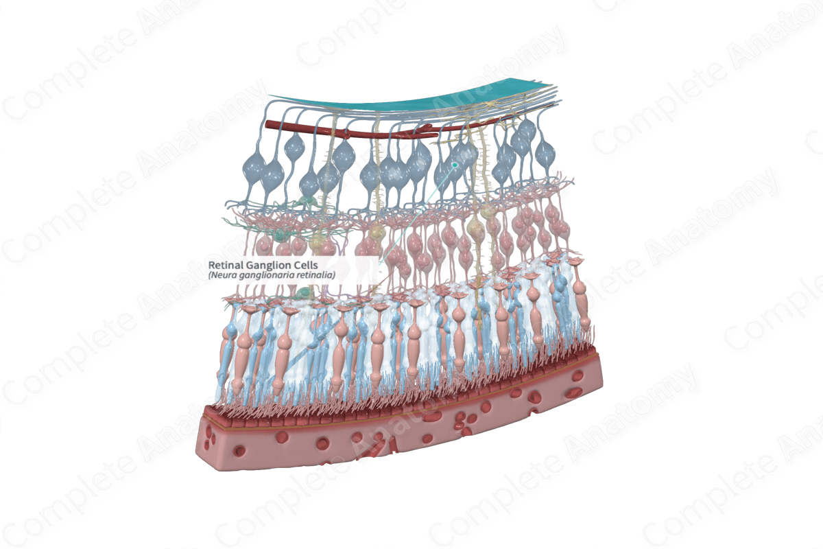
Structure and/or Key Features
Retinal ganglion cells are described as the third-order output neurons of the retina. There are approximately 0.7-1.5 million in the human retina. Retinal ganglion cells exhibit a topographic distribution across the retinal surface. They reach a peak density in a horizontally orientated elliptical ring surrounding the foveal center, though they are absent over the fovea itself (Standring, 2016; Ross and Pawlina, 2006). They are most numerous around in the perifoveal region of macula where they can be found to be stacked up to 10 cells thick. Their density remains high enough to be multiple cells thick in the macular region. Throughout most of the retina, they form a single layer with the distance between neighboring cells increasing with increasing eccentricity.
Retinal ganglion cell axons pass vitreal into a nerve fiber layer on the inner surface of the retina. These axons converge on the optic nerve head and proceed to exit the eye as the optic nerve. Axons in the human retina are typically non-myelinated, but are surrounded by the processes of radial glial cells and retinal astrocytes (Standring, 2016). Approximately 90% of the axons of retinal ganglion cells terminate within the lateral geniculate nucleus of the thalamus, while the remaining 10% project to subthalamic areas (Remington and Goodwin, 2011).
There is great morphological variation within the retinal ganglion cell population. Among the parameters on which ganglion cells vary are soma size, axon diameter (and consequent conduction velocity), polarity of primary dendrites (from bipolar, to multipolar), pattern and density of dendritic branching. Central projection target area of ganglion cells in the brain can also vary, thus making classification difficult (Remington and Goodwin, 2011).
Functional subsets of retinal ganglion cells have been identified, though the precise number of types and their function(s) is still being worked out. A popular method for the classification of retinal ganglion cells is by the layer in the lateral geniculate nucleus (LGN) in which they terminate. Located in the thalamus of the brain, the LGN is a relay center for the visual pathway that receives major sensory input from the retina. For example, a class of ganglion cells referred to as “P,” terminate in the parvocellular layers of the LGN. The most common P cell, known as the P1 or midget ganglion cell, has a small cell body with a single dendrite with two distinct levels of stratification of their terminal dendrites. P1 cells may only make contact with a single midget bipolar cell, which in turn only makes contact with a single cone cell forming a direct cone to bipolar to P1 ganglion cell channel that carries high contrast, fine detail and color information. Other ganglion cell types include P2 ganglion cells and M-type ganglion cells. P2 ganglion cells also terminate in the parvocellular layers of the LGN but have dense dendritic branching. M-cells project to the magnocellular layers of the LGN and possess a coarse and spiny dendritic tree (Remington and Goodwin, 2011). M-cells are also major contributors to the retinal input to the superior colliculus and other midbrain targets.
Anatomical Relations
Retinal ganglion cell bodies, along with displaced amacrine cells, retinal astrocytes, microglia, along with cells of the intraretinal vasculature are found in the ganglion cell layer of the retina. The dendritic processes of the ganglion cells make contact with bipolar and amacrine cells in the inner plexiform layer (Standring, 2016). The axons of ganglion cells pass into the layer of nerve fibers and converge at the optic disc to exit the eye as the optic nerve. Axonal fibers traveling from the medial retina approach the optic disc in a radial pattern. Those approaching the disc from the lateral, or temporal, side of the retina take an arcuate route. A bundle of fibers, known as the papillomacular fasciculus, is formed as axons from the macula travel directly toward the disc (Standring, 2016).
Function
Retinal ganglion cells receive visual signals from bipolar and amacrine cells. It carries these signals, via the optic nerve, to the visual centers of the brain, primarily the thalamic relay, lateral geniculate nucleus, midbrain, superior colliculus, and other subthalamic areas.
List of Clinical Correlates
- Glaucoma
- Neuroretinitis
- Devic’s disease
References
Dorland, W. (2011) Dorland's Illustrated Medical Dictionary. 32nd edn. Philadelphia, USA: Elsevier Saunders.
Ross, M. H. and Pawlina, W. (2006) Histology: A text and atlas. Lippincott Williams & Wilkins.
Standring, S. (2016) Gray's Anatomy: The Anatomical Basis of Clinical Practice. Gray's Anatomy Series 41 edn.: Elsevier Limited.
