Quick Facts
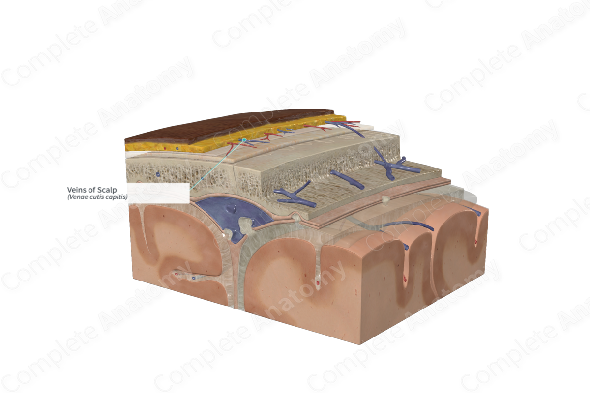
The veins of the scalp are located in the subcutaneous tissue and largely follow the course of the arteries.
Related parts of the anatomy
Structure
The scalp has a particularly rich blood supply, with vessels located in the dense subcutaneous tissue. The venous drainage of the anterior scalp is via the supraorbital and supratrochlear veins. The scalp anterior to the auricles is drained by the superficial temporal veins, while the scalp posterior to the auricles is drained by the posterior auricular veins. The occipital region of the scalp is drained by the occipital veins. The deep parts of the scalp in the temporal region are drained by the deep temporal veins (Moore, Dalley and Agur, 2013).
Key Features/Anatomical Relations
The veins that drain the scalp generally accompany the arteries. The supraorbital and supratrochlear veins terminate by forming the angular vein at the root of the nose. The superficial temporal veins terminate by joining the maxillary veins to form the retromandibular veins. The posterior auricular veins descend behind the auricle and unite with the posterior division of the retromandibular vein, thus forming the external jugular vein. The deep temporal veins are tributaries of the pterygoid venous plexus which is drained by the deep facial vein. Tributaries of the external jugular veins drain the scalp posteriorly and laterally.
Function
The veins of the scalp drain blood from the scalp.
References
Moore, K. L., Dalley, A. F. and Agur, A. M. R. (2013) Clinically Oriented Anatomy. Clinically Oriented Anatomy 7th edn.: Wolters Kluwer Health/Lippincott Williams & Wilkins.
