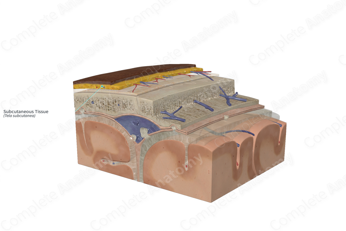
Quick Facts
The subcutaneous tissue is the layer of loose connective tissue situated just beneath the skin (Dorland, 2011).
Related parts of the anatomy
Structure
The subcutaneous tissue, or superficial fascia, of the scalp is composed of a dense connective tissue. It contains adipose tissue that is separated by fibrous septae. The thickness of the superficial fascia decreases with age (Brennan, Mahadevan and Evans, 2015).
The subcutaneous tissue is the most vascularized region of the scalp and contains many anastomosing arteries, veins, and lymphatic vessels. The vessels are attached to the fibrous connective tissue, thus, if a vessel is cut, the ends become retracted between the fibrous septae and may lead to profuse bleeding (Ellis and Mahadevan, 2014).
Key Features/Anatomical Relations
The subcutaneous tissue of the scalp lies below the skin to which it is firmly attached. It also attaches to the occipitofrontalis muscle and epicranial aponeurosis internally. Posteriorly, it is continuous with the subcutaneous tissue that covers the back of the neck. Laterally, the subcutaneous tissue of the scalp is continuous with the subcutaneous tissue overlying the temporal region.
Function
The subcutaneous tissue possesses fibers that join the skin of the scalp with the epicranial aponeurosis.
List of Clinical Correlates
—Scalp laceration
—Hematomas
References
Brennan, P. A., Mahadevan, V. and Evans, B. T. (2015) Clinical Head and Neck Anatomy for Surgeons. CRC Press.
Dorland, W. (2011) Dorland's Illustrated Medical Dictionary. 32nd edn. Philadelphia, USA: Elsevier Saunders.
Ellis, H. and Mahadevan, V. (2014) 'The surgical anatomy of the scalp', Surgery (Oxford), 32, pp. e1-e5.
