Quick Facts
Mitochondria are the small spherical to rod-shaped cytoplasmic organelles, consisting of inner and outer bilayer membranes with a space between them. The inner membrane is infolded to form a series of projections (cristae), and the space between the cristae is filled by the mitochondrial matrix, which contains DNA, RNA, ribosomes, and granules. Mitochondria generate energy (in the form of ATP synthesis) by the oxidation of nutrients, and they contain the enzymes of the tricarboxylic acid (Krebs) cycle and for fatty acid oxidation and oxidative phosphorylation. In response to toxic insults, they release enzymes that cause apoptosis. Mitochondria can replicate independently and code for the synthesis of some of their proteins; inheritance of mitochondrial DNA is maternal, and mitochondrial DNA defects cause a variety of diseases (Dorland, 2011).
Related parts of the anatomy
Structure
Mitochondria are small, membrane-bound, ovoid organelles approximately 0.5-2µm long. Each mitochondrion is lined by an inner and outer membrane. The outer membrane often attaches to other organelles, while the inner membrane is deeply folded to form deep invaginations, called cristae. The lumen of the mitochondria is surrounded by the inner membrane and contains the mitochondrial matrix (Gouspillou and Hepple, 2017).
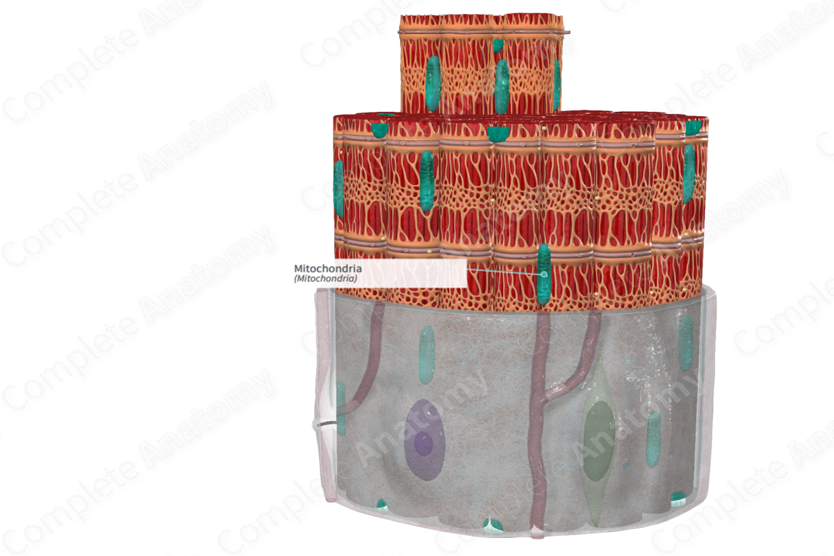
Key Features/Anatomical Relations
The majority of mitochondria are located between adjacent myofibrils and the sarcolemma.
Function
Mitochondria, along with liquid droplets and glycogen, are the primary source of metabolic support in the muscle fiber. Mitochondria provide chemical energy via the Krebs cycle, which results in the production of ATP from ADP. The mitochondrial matrix houses the enzymes required for this energy production. The number of mitochondria in a muscle fiber varies in response to sustained changes in activity.
List of Clinical Correlates
—Aging-related loss of muscle mass and function
—Disuse-induced muscle atrophy
—Ventilator-induced diaphragmatic dysfunction
—Duchenne and collagen muscular dystrophies
References
Dorland, W. (2011) Dorland's Illustrated Medical Dictionary. 32nd edn. Philadelphia, USA: Elsevier Saunders.
Gouspillou, G. and Hepple, R. T. (2017) Mitochondria in Skeletal Muscle Health, Aging and Diseases. Frontiers Research Topics.
