Quick Facts
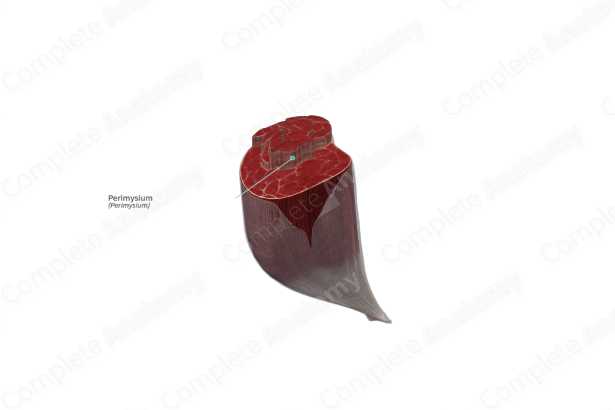
The perimysium is the connective tissue investing a fascicle of skeletal muscle fibers (Dorland, 2011).
Related parts of the anatomy
Structure
The perimysium is a tough and relatively thick layer of connective tissue that consists primarily of type I and type III collagen fibers. Some of the collagen fibers run alongside the underlying muscle fibers, while others are arranged in a “crisscross” pattern around the muscle fascicle.
Key Features/Anatomical Relations
The perimysium divides the skeletal muscle into various compartments/sections called fascicles, which contain bundles of muscle fibers. The three collagenous sheaths, the epimysium, perimysium, and endomysium, unite and fuse where the muscles connect to adjoining structures such as tendons. Underlying the perimysium proper is a network of loose irregularly arranged connective tissue which is also attached to the endomysium (MacIntosh, Gardiner and McComas, 2006).
Function
The perimysium is composed of collagen and elastin fibers, which contributes to the resistance of a muscle to tensile forces. The perimysium also provides a path for nerves and blood vessels, which innervate and supply the blood flow to the fascicles containing the muscle fibers (Martini et al., 2017).
List of Clinical Correlates
—Vasculitis
—Mysium damage
—Connective tissue disease
References
Dorland, W. (2011) Dorland's Illustrated Medical Dictionary. 32nd edn. Philadelphia, USA: Elsevier Saunders.
MacIntosh, B. R., Gardiner, P. F. and McComas, A. J. (2006) Skeletal Muscle: Form and Function. Human Kinetics.
Martini, F. H., Nath, J. L. and Bartholomew, E. F. (2017) Fundamentals of Anatomy & Physiology. Pearson Education.
