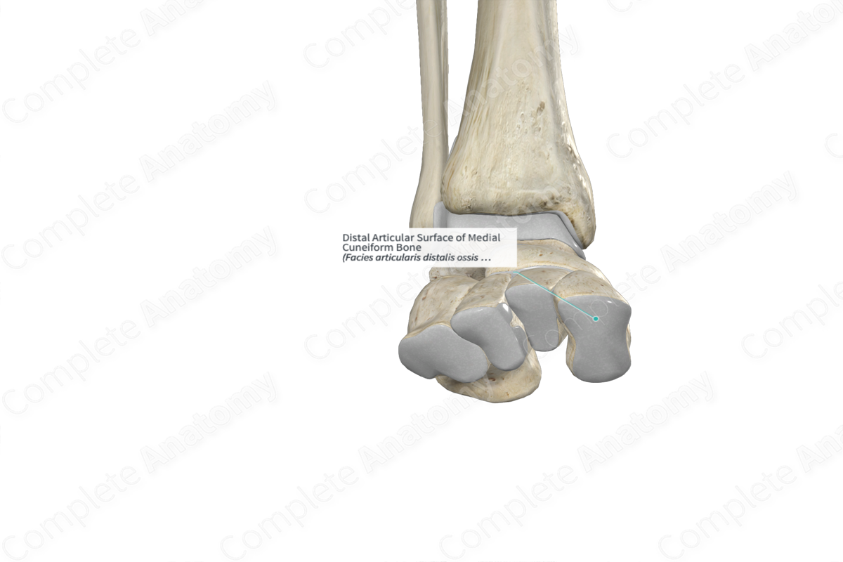
Distal Articular Surface of Medial Cuneiform Bone
Facies articularis distalis ossis cuneiformis medialis
Read moreDescription
The distal articular surface (anterior surface) is the kidney-shaped, anterior area of the medial cuneiform bone. It’s one of the six surfaces of the medial cuneiform bone, the other five being the plantar, dorsal, medial and lateral surfaces, and the proximal articular surface.
The distal articular surface articulates with the proximal articular facet of the first metatarsal bone, contributing to the formation of the tarsometatarsal joints.

Related parts of the anatomy
Learn more about this topic from other Elsevier products
Cuneiform Bone

The Lisfranc joint is formed of the three cuneiform bones and cuboid bone proximally with the five metatarsal bases distally linked by capsule-ligamentous structures.
