
Quick Facts
Location: Foot.
Bone Type: Short bone.
Key Features: Proximal and distal articular surfaces, plantar and dorsal surfaces, and medial and lateral cuneiform articular facets.
Articulates With: Navicular bone, lateral and medial cuneiform bones, second metatarsal bone.
Arterial Supply: Dorsalis pedis artery.


Related parts of the anatomy
Key Features & Anatomical Relations
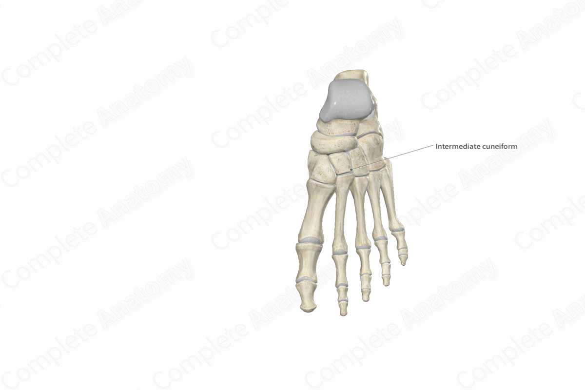
The intermediate (middle or second) cuneiform bone is one of the seven tarsal bones of the foot. It’s wedge-shaped and is found in the distal row of tarsal bones. The lateral cuneiform bone is classified as a short bone and includes the following bony features:
- surfaces: dorsal, plantar, medial and lateral surfaces, and proximal and distal articular surfaces;
- landmarks: medial and lateral cuneiform articular facets.
More information regarding these bony features can be found in the Surfaces and Landmarks tabs for this bone.
The intermediate cuneiform bone is located:
- proximal to the second metatarsal bone;
- distal to the navicular bone;
- medial to the lateral cuneiform bone;
- lateral to the medial cuneiform bone.
It articulates with the:
- second metatarsal bone, contributing to the formation of the tarsometatarsal joints;
- navicular bone, contributing to the formation of the cuneonavicular joint;
- medial and lateral cuneiform bones, contributing to the formation of the intercuneiform joints.
The intermediate cuneiform bone contributes to the formation of the medial longitudinal arch and transverse arch of the foot.
Ossification
Ossification of the intermediate cuneiform bone occurs at one ossification center, which appears within the third year (Standring, 2016).
Variations
In some individuals, an accessory ossicle may be associated with the intermediate cuneiform bone (Standring, 2016).
Surface Anatomy
With regard to surface anatomy, the intermediate cuneiform bone is difficult to palpate, as it’s more proximally positioned than the medial and lateral cuneiform bones.
List of Clinical Correlates
- Fracture of intermediate cuneiform bone
- Intercuneiform coalition
- Naviculocuneiform coalition
References
Standring, S. (2016) Gray's Anatomy: The Anatomical Basis of Clinical Practice. Gray's Anatomy Series 41st edn.: Elsevier Limited.
Learn more about this topic from other Elsevier products
Cuneiform Bone

The Lisfranc joint is formed of the three cuneiform bones and cuboid bone proximally with the five metatarsal bases distally linked by capsule-ligamentous structures.
