Quick Facts
Location: Foot.
Bone Type: Short bone.
Key Features: Proximal and distal articular surfaces, dorsal and plantar surfaces, second metatarsal articular facet, and intermediate cuneiform articular facet.
Articulates With: Navicular bone, first and second metatarsal bones, intermediate cuneiform bone.
Arterial Supply: Dorsalis pedis artery.


Related parts of the anatomy
Key Features & Anatomical Relations
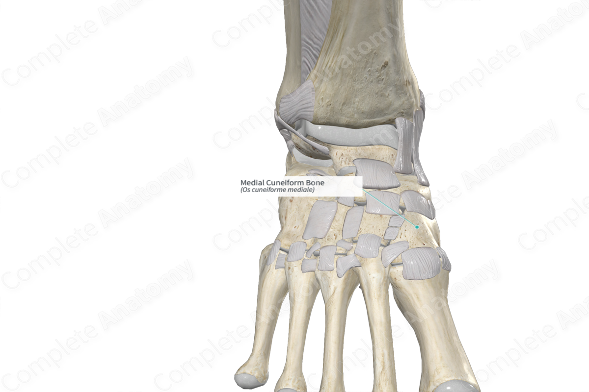
The medial (first) cuneiform bone is one of the seven tarsal bones of the foot. It's wedge-shaped and is found in the distal row of tarsal bones. The lateral cuneiform bone is classified as a short bone and includes the following bony features:
- surfaces: dorsal, plantar, medial and lateral surfaces, and proximal and distal articular surfaces;
- landmarks: second metatarsal articular facet and intermediate cuneiform articular facet.
More information regarding these bony features can be found in the Surfaces and Landmarks tabs for this bone.
The medial cuneiform bone is located:
- proximal to the first metatarsal bone;
- distal to the navicular bone;
- medial to the intermediate cuneiform bone and second metatarsal bone.
It articulates with the:
- first and second metatarsal bones, contributing to the formation of the tarsometatarsal joints;
- navicular bone, contributing to the formation of the cuneonavicular joint;
- intermediate cuneiform bone, contributing to the formation of the intercuneiform joints.
The medial cuneiform bone contributes to the formation of the medial longitudinal arch and transverse arch of the foot.
Ossification
Ossification of the medial cuneiform bone may occur at two ossification centers, which appear within the second year (Standring, 2016).
Variations
In some individuals:
- the medial cuneiform bone may be present in a bipartite condition (i.e. divided into two parts);
- an accessory bone may be associated with the medial cuneiform bone (Standring, 2016).
Surface Anatomy
With regard to surface anatomy, the medial surface of medial cuneiform bone can be palpated on the medial aspect of the foot, between the tuberosity of navicular bone and the base of first metatarsal bone.
List of Clinical Correlates
- Fracture of medial cuneiform bone
- Intercuneiform coalition
- Naviculocuneiform coalition
References
Standring, S. (2016) Gray's Anatomy: The Anatomical Basis of Clinical Practice. Gray's Anatomy Series 41st edn.: Elsevier Limited.
Learn more about this topic from other Elsevier products

