Quick Facts
Location: Foot.
Bone Type: Long bone.
Key Features: Head, body, base, medial and lateral surfaces, and proximal and distal articular facets.
Articulates With: Lateral cuneiform bone, second and fourth metatarsal bones, proximal phalanx of third toe.
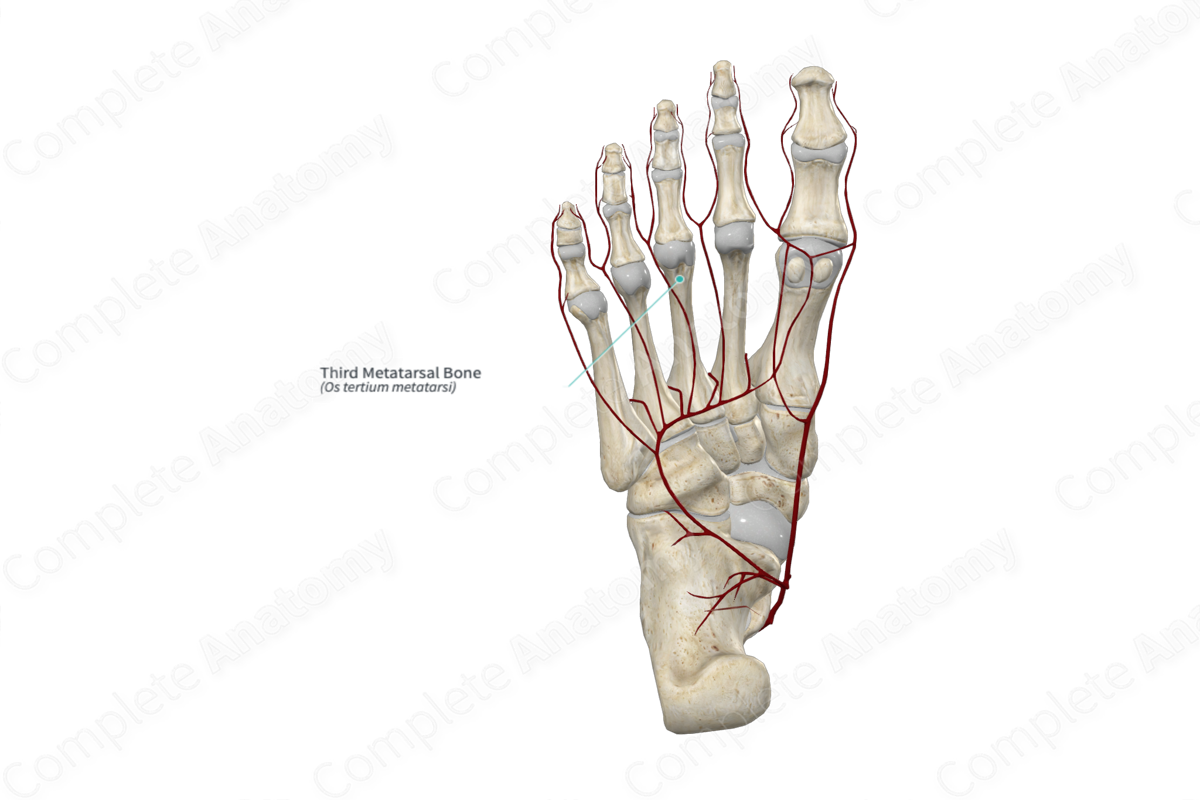
Arterial Supply: Plantar and dorsal metatarsal arteries.


Related parts of the anatomy
Key Features & Anatomical Relations
The third metatarsal bone is one of the five metatarsal bones of the foot. It is classified as a long bone and includes the following bony features:
- parts: head, body, and base;
- surfaces: medial and lateral surfaces;
- landmarks: proximal and distal articular facets, and second and fourth metatarsal articular facets.
More information regarding these bony features can be found in the Parts, Surfaces and Landmarks tabs for this bone.
The third metatarsal bone is located:
- proximal to the proximal phalanx of third toe;
- distal to the lateral cuneiform bone;
- medial to the fourth metatarsal bone;
- lateral to the second metatarsal bone.
It articulates with the:
- proximal phalanx of third toe at the third metatarsophalangeal joint;
- intermediate cuneiform bone, contributing to the formation of the tarsometatarsal joints;
- second and fourth metatarsal bones, contributing to the formation of the intermetatarsal joints.
Ossification
Ossification of the third metatarsal bone occurs at two ossification centers, these are found in the:
- body, which appears in utero at third month;
- head, which appears during the third to fourth years.
These ossification centers fuse with each other during the 17th and 20th years (Standring, 2016).
Surface Anatomy
The following bony features of the third metatarsal bone are relevant to surface anatomy:
- its dorsal aspect can be palpated;
- the head is palpable during plantarflexion of the toes.
List of Clinical Correlates
- Fracture
- Lisfranc injury
- Brachymetatarsia
- Neuropathic (diabetic) foot ulcer of the metatarsal head
References
Standring, S. (2016) Gray's Anatomy: The Anatomical Basis of Clinical Practice. Gray's Anatomy Series 41st edn.: Elsevier Limited.
Learn more about this topic from other Elsevier products
Third Metatarsal Bone

A fracture of the lateral, or more rarely the medial, condyle of the third metacarpal or metatarsal bone results in acute-onset, severe lameness.
