Quick Facts
Location: Vertebral column.
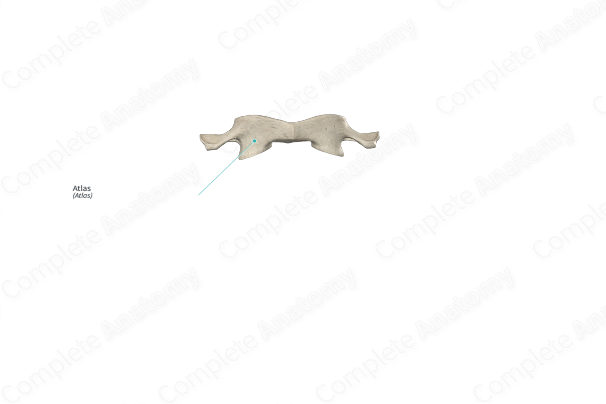
Bone Type: Irregular bone.
Key Features: Lateral masses, anterior and posterior arches, and transverse processes.
Articulates With: Occipital bone and axis.
Arterial Supply: Vertebral arteries.


Related parts of the anatomy
Key Features & Anatomical Relations
The atlas (first cervical vertebra or vertebra C1) is one of the seven cervical vertebrae of the vertebral column. It is considered an atypical cervical vertebra because some of its features differ to those of typical cervical vertebrae, such as the absence of a vertebral body and spinous process. The atlas is classified as an irregular bone, and it includes the following bony features:
- parts: lateral masses, anterior and posterior arches, and transverse processes;
- landmarks: superior and inferior articular facets, anterior and posterior tubercles, grooves for vertebral arteries, and transverse ligament tubercles.
More information regarding these and other bony features can be found in the Parts and Landmarks tabs for this bone.
The atlas is located:
- superior to the axis (second cervical vertebra);
- inferior to the occipital bone.
It articulates with the:
- axis at both the median and lateral atlantoaxial joints;
- occipital bone at the atlantooccipital joints.
Ossification
Ossification of the atlas occurs at three ossification centers, these are found in the:
- right and left lateral masses, with one center found in each, which appear in utero during the second month;
- anterior arch, which appears during the first year after birth.
The ossification centers for the right and left lateral masses fuse with each other to form the posterior arch during the third to fourth years. The anterior arch fuses with the lateral masses during the sixth to eighth years (Standring, 2016).
Variations
In some individuals:
- the posterior arch of atlas may be present as incompletely fused;
- the superior articular facets may be present in a bipartite condition (i.e., divided into two parts);
- the atlas may be fused with the occipital bone, known as atlantooccipital assimilation (Tubbs, Shoja and Loukas, 2016).
Surface Anatomy
The posterior tubercle of the atlas is difficult to palpate, as it lies deep to the nuchal ligament.
List of Clinical Correlates
- Fracture
- Osteoporosis
- Spinal stenosis
- Scoliosis
- Atlantooccipital assimilation
- Dislocation of atlantooccipital joints
- Dislocation of atlantoaxial joints
- Spina bifida
References
Standring, S. (2016) Gray's Anatomy: The Anatomical Basis of Clinical Practice. Gray's Anatomy Series 41st edn.: Elsevier Limited.
Tubbs, R. S., Shoja, M. M. and Loukas, M. (2016) Bergman's Comprehensive Encyclopedia of Human Anatomic Variation. Wiley.
