Quick Facts
Location: Thoracic cage.
Bone Type: Flat bone.
Key Features: Head, neck, tubercle, body, angle, and costal groove.
Articulates With: Fifth and sixth thoracic vertebrae, sixth costal cartilage.
Arterial Supply: Anterior intercostal branches of internal thoracic and posterior intercostal arteries.


Related parts of the anatomy
Key Features & Anatomical Relations
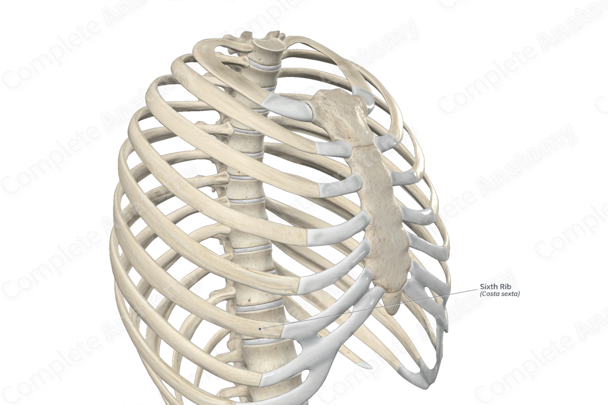
The sixth rib is one of the seven true ribs of the thoracic cage. It is considered a typical rib because it consists of a head with two articular facets, a neck, a tubercle, and a body. It does not have any extra bony features.
The sixth rib is classified as a flat bone and includes the following bony features:
- parts: head, neck, tubercle, body, and costal end;
- surfaces: internal and external surfaces, and superior and inferior borders;
- landmarks: angle, costal groove, crests on the head and neck, and articular facets on the head and tubercle.
More information regarding these and other bony features can be found in the Parts, Surfaces, and Landmarks tabs for this bone.
The sixth rib is located:
- superior to the seventh rib;
- inferior to fifth rib;
- lateral to the sixth costal cartilage and fifth and sixth thoracic vertebrae.
It articulates with the:
- sixth costal cartilage at the sixth costochondral joint;
- fifth and sixth thoracic vertebrae at the sixth costovertebral joint.
Ossification
Ossification of the sixth rib occurs at ossification centers found in the:
- body, which appears in utero during the second month;
- head, which appears during puberty;
- tubercle, which appears during puberty.
The ossification centers for the head and tubercle fuse with the body of the sixth rib within the fourteenth to twentieth years (Cunningham, Scheuer and Black, 2016).
Variations
In some individuals:
- the sixth rib may be fused with adjacent ribs;
- the costal end of the sixth rib may be bifid in appearance (Tubbs, Shoja and Loukas, 2016).
Surface Anatomy
The sixth rib is easily palpated and is located by palpating four ribs down from the second rib.
List of Clinical Correlates
- Fracture of sixth rib
- Flail chest
- Poland syndrome
- Asphyxiating thoracic dysplasia/Jeune syndrome
References
Cunningham, C., Scheuer, L. and Black, S. (2016) Developmental Juvenile Osteology. Elsevier Science.
Tubbs, R. S., Shoja, M. M. and Loukas, M. (2016) Bergman's Comprehensive Encyclopedia of Human Anatomic Variation. Wiley.
Learn more about this topic from other Elsevier products
Rib Cage

The rib cage forms the bony margins of the chest wall and is composed of the ribs, costal cartilages, sternum and thoracic vertebrae.
