Quick Facts
Location: Vertebral column.
Bone Type: Irregular bone.
Key Features: Vertebral body, laminae, pedicles, superior and inferior articular processes, and transverse and spinous processes.
Articulates With: Axis and fourth cervical vertebra.
Arterial Supply: Vertebral arteries.


Key Features & Anatomical Relations
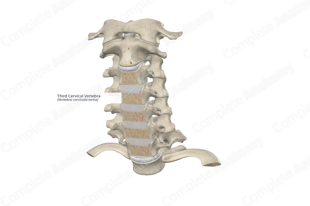
The third cervical vertebra (vertebra C3) is one of the seven cervical vertebrae of the vertebral column. It is considered a typical cervical vertebra because it has similar characteristic features to those of the fourth, fifth and sixth cervical vertebrae. The third cervical vertebra is classified as an irregular bone, and it includes the following bony features:
- parts: vertebral body, laminae, pedicles, superior and inferior articular processes, and transverse and spinous processes;
- surfaces: superior and inferior intervertebral surfaces, superior and inferior annular epiphyses, and vertebral arch;
- landmarks: superior and inferior vertebral notches, anterior and posterior tubercles, uncinate processes, and superior and inferior articular facets.
More information regarding these and other bony features can be found in the Parts, Surfaces, and Landmarks tabs for this bone.
The third cervical vertebra is located:
- superior to the fourth cervical vertebra;
- inferior to the axis (second cervical vertebra).
It articulates with the axis and fourth cervical vertebra at the intervertebral symphyses and zygapophyseal joints.
Ossification
Ossification of all typical cervical vertebrae occurs at eight ossification centers, these are found in the:
- vertebral body, which appears in utero during the second to fourth months;
- right and left halves of the vertebral arch, with one center found in each, which appear in utero during the third month;
- right and left transverse processes, with one center found in each, which appear during puberty;
- spinous process, which appears during puberty;
- superior and inferior annular epiphyses, with one center found in each, which appear during puberty.
The ossification centers for the right and left halves of the vertebral arch fuse with each other during the first year after birth. The vertebral arch fuses with the vertebral body during the third year. The remaining centers fuse with the vertebral arch and body during early adulthood (Standring, 2016).
Variations
In some individuals:
- accessory transverse foramina may be present;
- the spinous process may be present in a nonbifid form (Tubbs, Shoja and Loukas, 2016).
Surface Anatomy
The spinous process of the third cervical vertebra is difficult to palpate, as it lies deep to the nuchal ligament.
List of Clinical Correlates
- Fracture
- Osteoporosis
- Spinal stenosis
- Scoliosis
- Dislocation
- Spina bifida
- Block vertebra
References
Standring, S. (2016) Gray's Anatomy: The Anatomical Basis of Clinical Practice. Gray's Anatomy Series 41st edn.: Elsevier Limited.
Tubbs, R. S., Shoja, M. M. and Loukas, M. (2016) Bergman's Comprehensive Encyclopedia of Human Anatomic Variation. Wiley.
