Quick Facts
Compact bone is bone substance that is dense and hard (Dorland, 2011).
Structure/Morphology
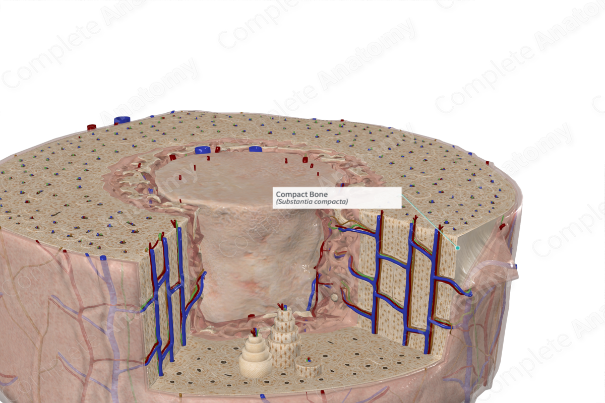
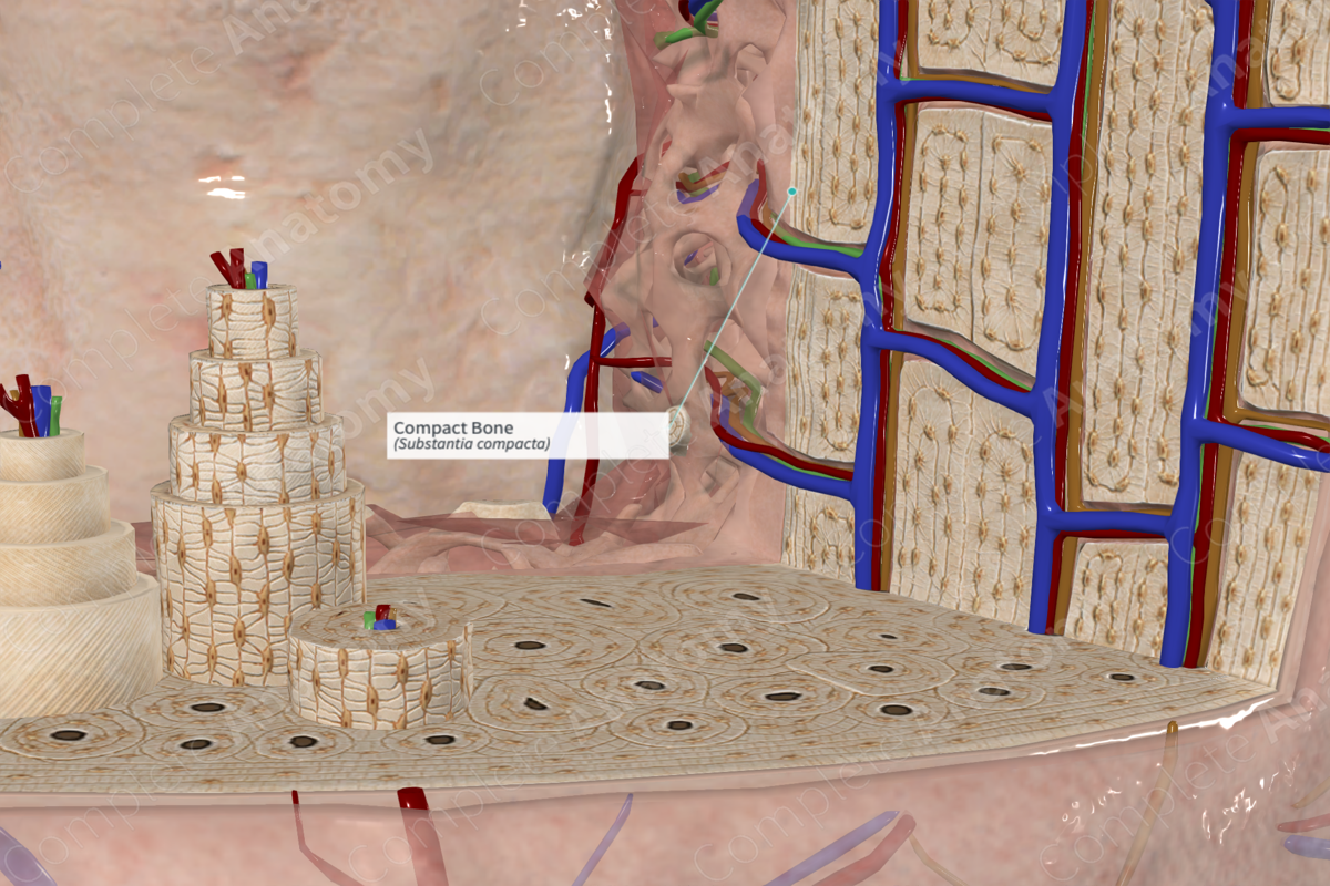
Compact bone forms the outer dense covering of bone tissue. Approximately 80% of bone in the adult human skeleton is composed of compact bone (Draper and Marshall, 2014). Compact bone is comprised of functional units called osteons. In dense, compact regions of long bones, osteons lie parallel to each other and are orientated with the long axis of the bone itself and appear round in cross-section.
In compact bone, osteons are identified by their central, nutrient canal surrounded by 5–20 concentric rings of lamellar bone (Standring, 2016). The bony matrix of the lamellar compact bone is dense and only small spaces called lacunae are found. Interspersed between the osteons of compact bone are nonuniform spaces comprised of interstitial lamellae. The interstitial lamellae have a haphazard orientation which is in contrast to the circumferential lamellae seen surrounding the nutrient canals within the osteons. Inner and outer circumferential lamellar layers are found lining the internal and external surfaces of compact bone.
Anatomical Relations
The outer circumferential lamellar layer of cortical bone is surrounded by the osteogenic periosteal layer of the periosteum. The innermost portion of the cortical bone is made up of the inner circumferential lamella. This portion of the inner cortical surface is adjacent to the spongy bone. The spongy bone, with its irregular trabeculae, does not completely cover the internal surface of the cortical bone and is lined by an endosteum.
Function
The arrangement of compact, lamellar bone confers the strength required for structural support to the body. It assists in movement by giving leverage to muscles and by acting as a rigid support for muscle contraction. Compact bone provides structural support for the body and protects internal structures (e.g., the heart and the spinal cord). It also acts as a reservoir for important ions such as calcium and phosphate. The release of these ions is very tightly balanced to preserve mineral homeostasis (Standring, 2016).
References
Dorland, W. (2011) Dorland's Illustrated Medical Dictionary. 32nd edn. Philadelphia, USA: Elsevier Saunders.
Draper, N. and Marshall, H. (2014) Exercise Physiology: For Health and Sports Performance. Taylor & Francis.
Standring, S. (2016) Gray's Anatomy: The Anatomical Basis of Clinical Practice. Gray's Anatomy Series: Elsevier Limited.

