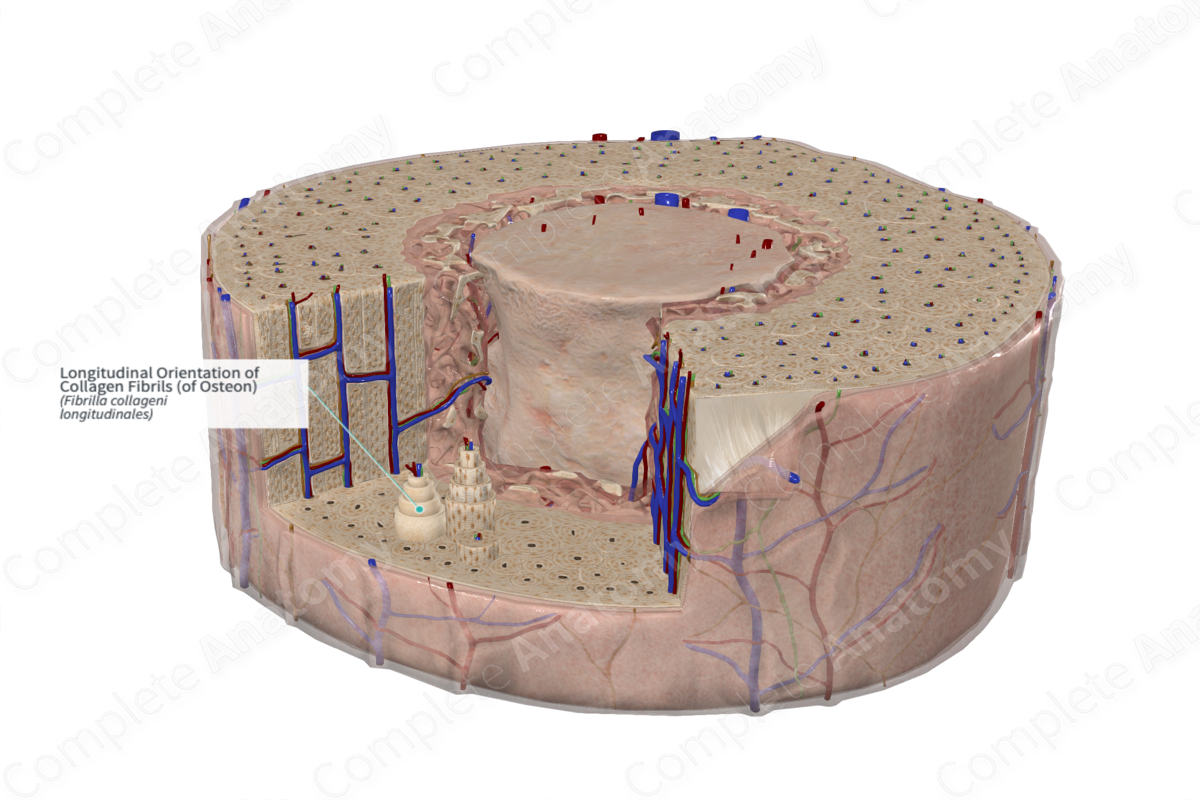
Longitudinal Orientation of Collagen Fibrils (of Osteon)
Fibrilla collageni longitudinales
Read moreQuick Facts
Within a single osteon concentric lamella, collagen fibers are arranged longitudinally. Fibers in adjacent lamellae alternate in orientation giving the lamellae a helical appearance (Standring, 2016).
Structure/Morphology
Longitudinally oriented collagen fibrils are arranged parallel to one another within a single lamella. Between adjacent lamellae, fibrils alternate in orientation. This gives the lamellae a helical pattern, akin to plywood when cut in cross-section. These fibrils are mainly collagen type I, but they have stronger internal cross-linkage between fibrils compared to other connective tissues (Standring, 2016).
There are a variety of orientations that the collagen fibers can take within the bone, including transverse, alternating and longitudinal (Spiesz, Kaminsky and Zysset, 2011). Collagen fibers oriented longitudinally are most common in the long bones, where there is high tensile stress. Transverse collagen fibrils are generally found in compact bone that is exposed to high compression stress.
Anatomical Relations
Collagen fibrils are a key component of the bone matrix and account for approximately one third of all extracellular matrices in bone tissue. The space between collagen fibrils is larger than those in other connective tissues to facilitate mineral deposition during bone formation.
Function
The stability of the collagen fibrils and the space between the fibrils aid in the strength and deposition of new bone formation and help confer structural support. In addition, the alternating collagen fibril orientations between different lamellae also help facilitate a greater strength (Standring, 2016).
References
Spiesz, E. M., Kaminsky, W. and Zysset, P. K. (2011) 'A quantitative collagen fibers orientation assessment using birefringence measurements: Calibration and application to human osteons', Journal of Structural Biology, 176(3), pp. 302-306.
Standring, S. (2016) Gray's Anatomy: The Anatomical Basis of Clinical Practice. Gray's Anatomy Series: Elsevier Limited.
