Quick Facts
There is an extensive network of periosteal vessels along the entire length of a bone shaft. They are capable of supplying up to one third of the outer compact bone.
Related parts of the anatomy
Structure/Morphology
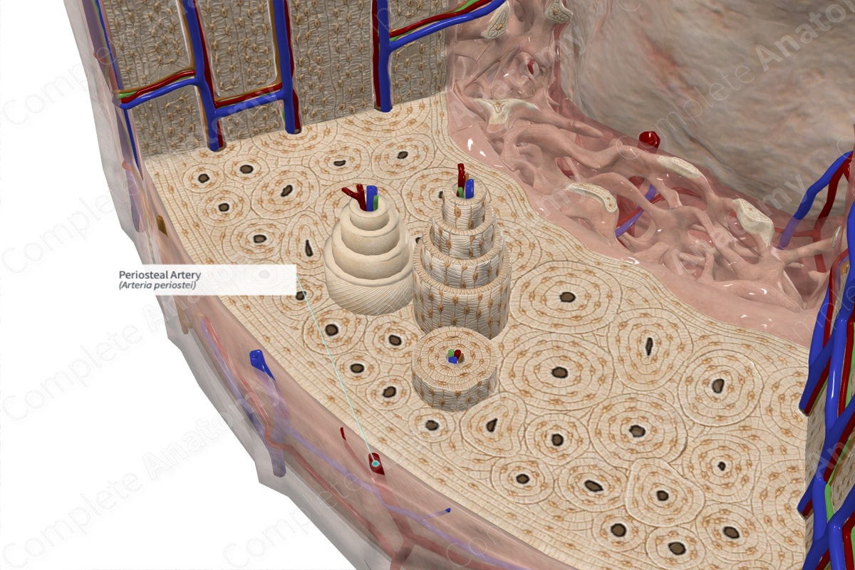
Osseous blood flow is described as centrifugal (radiating from the central medullary cavity towards the external cortical bone via the nutritional artery) or centripetal (radiating inward from the external periosteum via periosteal arterioles). The centripetal flow accounts for the flow of blood from the periosteum into the outer one third of cortical bone. These vessels enter the outer one third of the cortex and anastomose with the medullary system within the perforating canals. They lie in their respective perforating canals and anastomose with their medullary counterparts to form a rich microvascular network inside cortical bone (Buck and Dumanian, 2012).
Anatomical Relations
The periosteum has an intrinsic network of vessels that are derived from adjacent soft tissues such as the musculoperiosteal and fascicoperiosteal plexus from nearby muscles and fascial planes. These vessels form an arteriole network and provide intrinsic blood supply to the periosteum. This network lies between the two periosteal layers. The terminal branches of the intrinsic periosteal arteriole network extend directly into the underlying cortical bone.
Function
The periosteal artery supplies blood to the periosteum and outer one third of the cortex. In the central canals of the external osteons, the periosteal arteries anastomose with the medullary arteries, forming a periosteocortical anastomosis. This anastomosis represents a direct connection between periosteal blood and blood from the nutrient foramina. This is pertinent for the survival of the outer cortex when medullary blood supply is diminished or blocked.
References
Buck, D. W., 2nd and Dumanian, G. A. (2012) 'Bone biology and physiology: Part I. The fundamentals', Plast Reconstr Surg., 129(6), pp. 1529-4242.
