Quick Facts
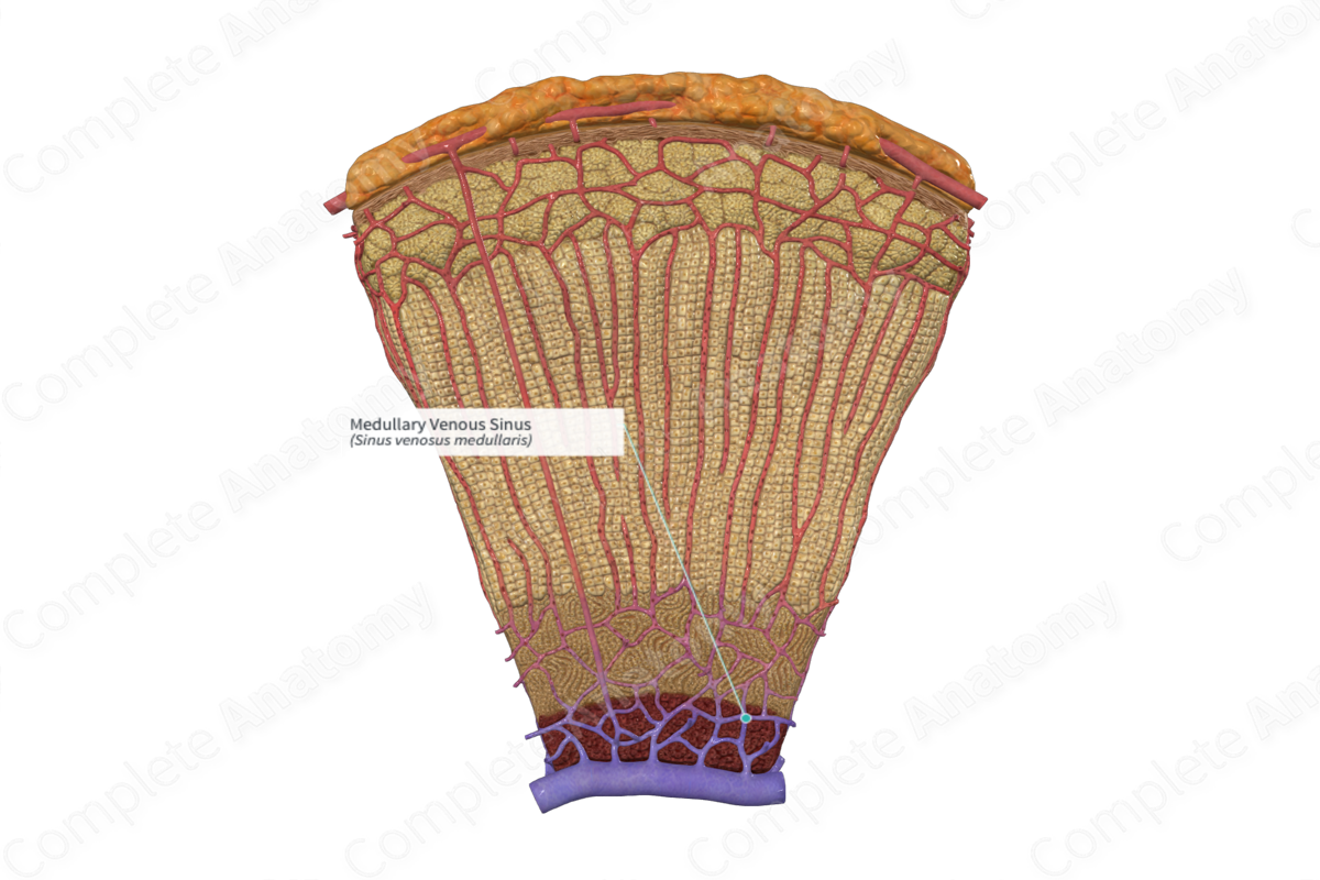
The medullary venous sinus is formed by the convergence of medullary cortical sinusoids and the medullary artery. They merge with the central veins.
Related parts of the anatomy
Structure and/or Key Features:
The medullary venous sinus is formed by the convergence of medullary cortical sinusoids and the medullary artery. The venous sinuses pass among the chromaffin cells in the medullary tissue. They go on to merge with the central veins, carrying the secreted hormones in the blood from the various suprarenal regions. At the hilum, the central veins emerge as the left and right suprarenal veins, which empty into the left renal vein on the left, and the inferior vena cava on the right (Inomata and Sasano, 2015).
Function
The medullary venous sinus transports the secreted hormones of the suprarenal gland to the systemic circulation.
References
Inomata, A. & Sasano, H. (2015) Practical approaches for evaluating adrenal toxicity in nonclinical safety assessment. J Toxicol Pathol, 28(3), 125-32.
