Quick Facts
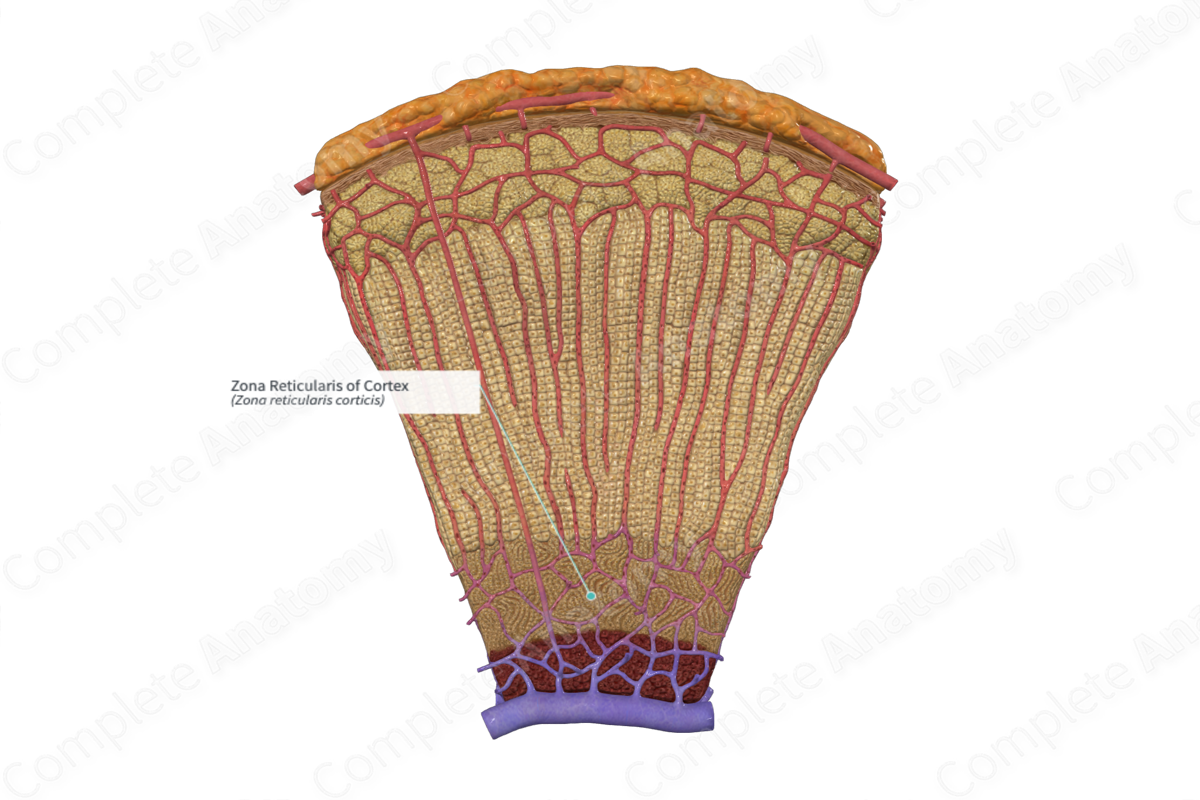
The zona reticularis is the inner layer of the suprarenal cortex, consisting of cells arranged as clearly anastomosing cords, and abutting the medulla (Dorland, 2011).
Related parts of the anatomy
Structure and/or Key Features:
The cortex of the suprarenal gland is comprised of three distinct morphological zones, the zona glomerulosa, zona fasciculata, and the zona reticularis. The zona reticularis is the innermost section of the suprarenal cortex. Unlike the other cortical regions, it does not differentiate completely until childhood.
The zona reticularis is made of a network of cellular pillars interconnected with each other. The cells of this region contain numerous lysosomes to generate a cholesterol-rich supply. This lysosomal digestion process results in speckles of yellow-brown pigments within the cell (Pocock, Richards and Richards, 2013).
Anatomical Relations:
The zona reticularis is the innermost layer of the suprarenal cortex and is contained between the zona fasciculata on the outside and the suprarenal medulla on the inside.
The long arterial sinusoids from the zona fasciculata form a fenestrated network of vessels to supply the zona reticularis tissue.
Function
The primary function of the cortex is to produce steroid hormones. In particular, the zona reticularis secretes androgens, such as dehydroepiandrosterone (DHEA), its sulfated form DHEAS, and androstenedione. These are prohormones which must be converted to sex hormones in the gonads. In females, they are thought to contribute to the development of secondary sexual characteristics (Lois et al, 2014).
Clinical Correlates
—Precocious puberty
—Congenital suprarenal hyperplasia
References
Dorland, W. (2011) Dorland's Illustrated Medical Dictionary. 32nd edn. Philadelphia, USA: Elsevier Saunders.
Lois, K., Kassi, E., Prokopiou, M. & Chrousos, G. P. (2014) Adrenal Androgens and Aging, 2014. Available online
Pocock, G., Richards, C. D. & Richards, D. A. (2013) Human Physiology, 4 edition. OUP Oxford.
