Quick Facts
The medulla of suprarenal gland is the inner, reddish brown, soft part of the suprarenal gland; it synthesizes, stores, and releases catecholamines (Dorland, 2011).
Structure and/or Key Features
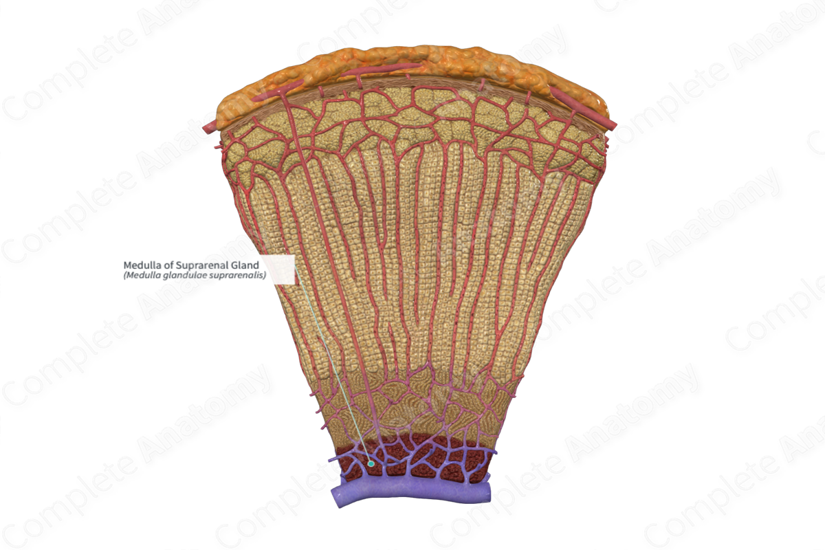
The medulla of the suprarenal gland forms the inner layer of the gland. There are no different morphological zones within the tissue, unlike the cortex of the suprarenal gland. The medulla is dark red/gray and is derived from neuroectoderm.
The medulla comprised of small assemblies and pillars of chromaffin cells (pheochromocytes) which line the venous sinuses. These chromaffin cells are part of the neuroendocrine system and are comparable to postganglionic sympathetic neurons. They synthesize, store, and secrete catecholamines into the bloodstream. Storage occurs by packing the catecholamines into granules by chromogranin proteins. Essentially, the medulla is a large sympathetic ganglion, which releases catecholamines into circulation. Secretion of medullary catecholamines occurs largely in response to sympathetic stimulation via splanchnic nerve activity, and a variety of physiological and emotional stresses.
The medulla has a dual blood supply. It is supplied by arterial sinusoids which perfuse the suprarenal cortex. It is also supplied by direct branches from the capsular plexus, the medullary arteries, penetrating deep into the medullary tissue without branching. The medulla is drained by medullary veins, which form the suprarenal veins at the hilum of the suprarenal gland. Lymph is drained to the hilum, where lymph vessels transport lymph to the lateral aortic lymph nodes.
Anatomical Relations:
The suprarenal medulla forms the inner part of the suprarenal gland. It is subjacent to the suprarenal cortex.
Function
The primary function of the suprarenal medulla is the secretion of catecholamines into the venous sinuses. They have a variety of bodily functions, including increasing heart rate and blood pressure. Of note, norepinephrine increases diastolic pressure, while epinephrine reduces the diastolic pressure on account of its vasodilatory effects within skeletal muscle.
The mechanism of releases is exocytosis of granule-containing catecholamines in response to depolarization of the chromaffin cells via sympathetic activation. Epinephrine is the majority of secretions (around 80%), with norepinephrine being the minority (around 20%). The release of the catecholamines is mediated by splanchnic nerve activity. They are also secreted in response to physiological stresses such as hypoglycemia, hypotension, hemorrhage, and to emotional stresses including fear, anger, and sexual arousal.
Clinical Correlates
—Pheochromocytoma
—Von Hippel-Lindau disease
References
Dorland, W. (2011) Dorland's Illustrated Medical Dictionary. 32nd edn. Philadelphia, USA: Elsevier Saunders.
