Quick Facts
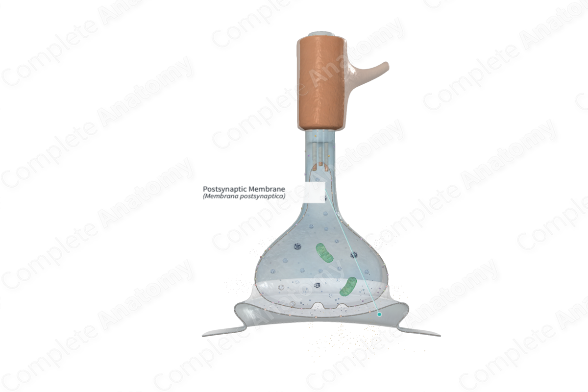
The postsynaptic membrane is the area of plasma membrane of a postsynaptic cell, either a muscle fiber or a neuron, that is within the synapse and has areas especially adapted for receiving neurotransmitters (Dorland, 2011).
Related parts of the anatomy
Structure
The postsynaptic membrane is the site at which the neurotransmitter will interact with a receptor. Part of the plasmalemma of the postsynaptic neuron exhibits dense material. This postsynaptic density contains a complex arrangement of proteins that translate the neurotransmitter-receptor interaction into an intracellular signal, traffic and anchor neurotransmitter receptors to the postsynaptic membrane, and some proteins modulate the activity of the receptors.
Neurotransmitters diffuse across the synaptic cleft and act on multiple receptors. They act either on:
—metabotropic receptors to activate a G-protein signaling cascade;
—ionotropic receptors to open the membrane ion channels.
Metabotropic channels bind a specific neurotransmitter and interact with G-protein on the intracellular domain. G-protein signals the synthesis of a second messenger. Metabotropic receptors are assoaciated with modulation of neuronal activity.
Ionotropic receptors contain integral transmembrane ion channels called transmitter or ligand-gated channels. If a neurotransmitter binds to ionotropic receptors, the channel opens to allow selective movement of ions in and out of the cell and an action potential is generated in the effector cell. This type of signaling is very fast and occurs in somatic motor pathways in the peripheral nervous system and in major neuronal pathways in the brain .
Depending on which receptors they interact with, various neurotransmitters are capable of generating a diverse range of excitatory or inhibitory postsynaptic actions. The consequent generation of a nerve impulse in a postsynaptic neuron is highly dependent on the addition of both the excitatory and inhibitory neuronal impulses reaching the postsynaptic neuron.
With excitatory synapses, the release of acetylcholine, glutamine, or serotonin neurotransmitters open transmitter-gated sodium channels (or other cation channels). This results in the influx of sodium into the cell which initiates a local voltage reversal (depolarization) of the postsynaptic membrane, leading to the generation of an action potential and a nerve impulse.
With inhibitory synapses, the release of γ-aminobutyric acid (GABA) or glycine neurotransmitters open transmitter-gated chloride channels (or other anion channels). This results in the influx of chloride into the cell, leading to hyperpolarization of the postsynaptic membrane. This process renders the postsynaptic membrane more negative, resulting in a more challenging generation of action potential.
Whether a nerve impulse is generated in a postsynaptic neuron depends on input of excitatory or inhibitory actions from hundreds of synapses on the postsynaptic neuron.
Neurotransmitters bind to their appropriate receptor for only about one millisecond before dissociating from it. If the presynaptic cell continues releasing neurotransmitter then one molecule will replace the other on the receptor and the postsynaptic cell will be restimulated (Ross and Pawlina, 2006).
Anatomical Relations
The postsynaptic region is a part of the distal neuron in a chemical synapse. The postsynaptic region is separated from the presynaptic region by the synaptic cleft.
Function
The postsynaptic membrane is vital in the process of neurotransmission. It has a number of receptors that are coupled to ion channels within its structure. These receptors are the sites on which neurotransmitters from the presynaptic element bind and act on, opening the ion channels to allow the influx of specific ion channels, to exert excitatory or inhibitory effects that either depolarize or hyperpolarize the postsynaptic neuron (Ross and Pawlina, 2006).
Clinical Correlates
—Drugs are being developed now to modify synaptic transmission by interfering with the processes of transmission at the postsynaptic membrane (Splittgerber, 2018).
—Myasthenia gravis is an autoimmune disorder, where antibodies are produced against nicotinic acetylcholine receptors on the postsynaptic membrane.
—In high concentrations, nicotine acts as a blocking agent by stimulating the postganglionic neuron causing and maintaining depolarization such that the postganglionic neuron will fail to respond to any stimulant.
References
Dorland, W. (2011) Dorland's Illustrated Medical Dictionary. 32nd edn. Philadelphia, USA: Elsevier Saunders.
Ross, M. H. and Pawlina, W. (2006) Histology: A text and atlas. Lippincott Williams & Wilkins.
Splittgerber, R. (2018) Snell's Clinical Neuroanatomy. Wolters Kluwer Health.
