Quick Facts
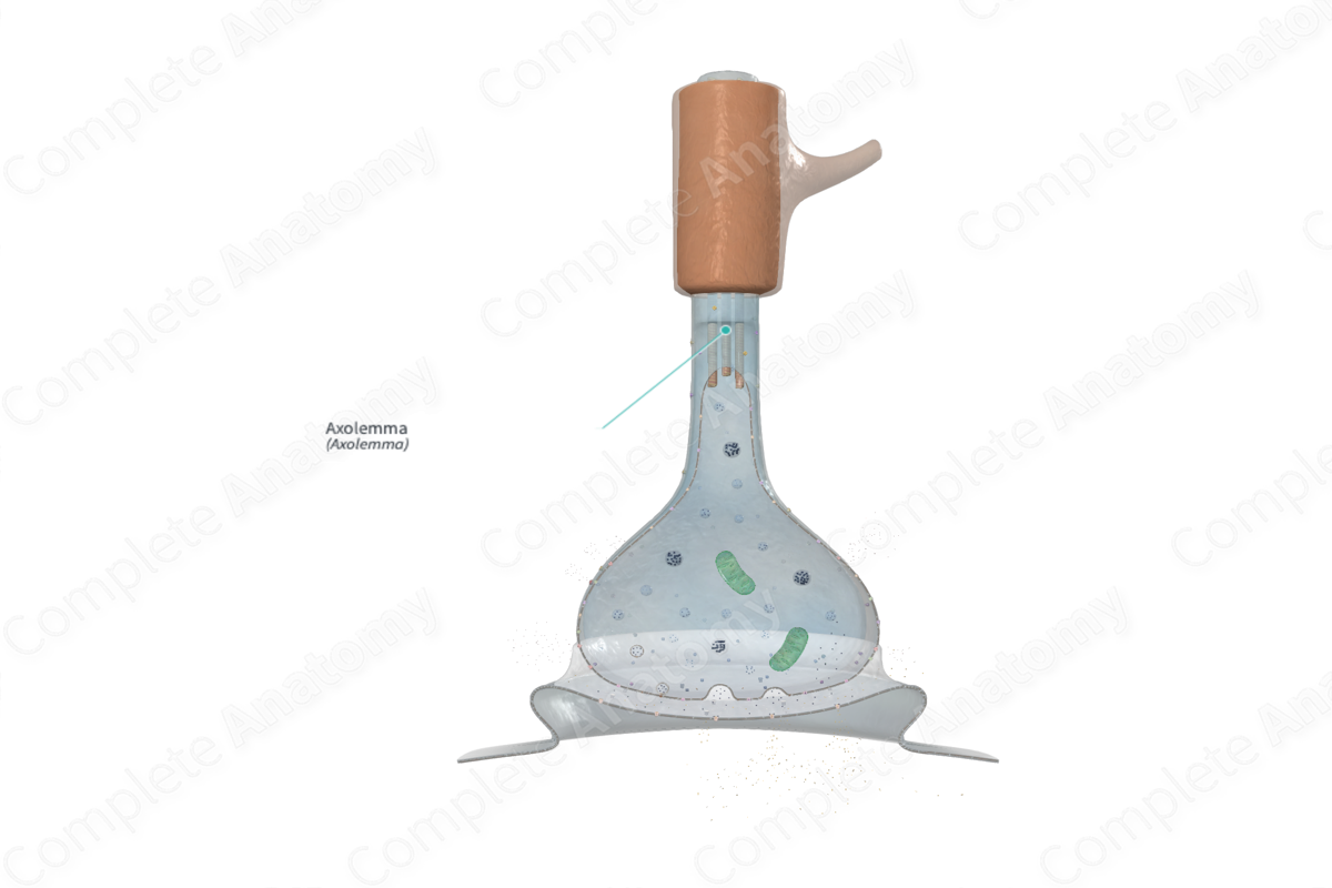
The axolemma is the plasma membrane of an axon (Dorland, 2011).

Structure
An axon is bound by plasmalemma (or plasma membrane) known as the axolemma. The axolemma is composed of a lipid bilayer and a submembrane skeleton, which is formed by the protein spectrin and actin, and are linked by ankyrin and a number of other constituents of the transmembrane proteins. This skeletal framework of spectrin and ankyrin aid in the anchoring of voltage-gated channels to the axolemma.
The propagation of nerve impulses depends on the structure of the axolemma, concentrations of electrolytes in the axolemma and the extracellular space, and selective permeability of the axolemma to the specific ions. The nerve signals are conveyed using action potentials that are generated with alterations to the permeability of the axolemma to sodium and potassium ions (Ross and Pawlina, 2006).
Anatomical Relations
The axolemma is the plasma membrane of the axonal process.
Function
The axolemma possesses various transmembrane channels which aid neurotransmission by enabling the passage of various ions, a process which initiates and propagates action potentials (Ross and Pawlina, 2006).
Clinical Correlates
—Diffuse axonal injury is characterized by disruption of the axon cytoskeleton.
—Stretching of axons during traumatic injury causes physical disruption to, and proteolytic degradation of, the cytoskeleton.
References
Dorland, W. (2011) Dorland's Illustrated Medical Dictionary. 32nd edn. Philadelphia, USA: Elsevier Saunders.
Ross, M. H. and Pawlina, W. (2006) Histology: A text and atlas. Lippincott Williams & Wilkins.
