Voltage-Gated Sodium Channel
Quick Facts
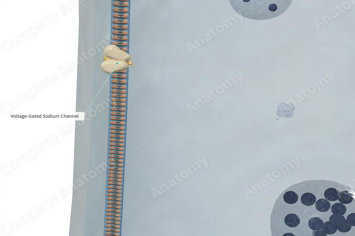
A sodium channel is a voltage-gated channel selective for the passage of sodium ions; sodium channels are activated by membrane depolarization and are the main causes of depolarization and repolarization of the nerve membrane during the propagation of action potentials (Dorland, 2011).
Related parts of the anatomy
Structure
The voltage-gated sodium channel is created by a single long polypeptide resulting in “pores” in the axolemma that are highly selective to Na+. At a negative resting membrane potential, the pore remains closed. When the membrane is depolarized to threshold, the molecule reconfigures such that Na+ can pass through the pore.
Voltage-gated sodium ion channels can exist in three main conformational states: closed, open, and inactivated. They change from one conformational state to the other on neurotransmitter binding.
Prior to the generation of an action potential, the membrane is at its resting potential and the voltage-gated sodium channels are in their deactivated conformational state. When the membrane potential reaches -55 mV approximately, a conformational change occurs in the sodium ion channel protein and “opens” the channel to allow the influx of sodium ions. This results in the depolarization of the cell and an initiation of the upstroke of the action potential. This step is the “activation” step. At this point, the voltage-gated sodium channel is referred to as being open and active (Ross and Pawlina, 2006; Bear, Connors and Paradiso, 2007).
Following the opening and activation of the voltage-gated sodium channel, influx of sodium ions, and subsequent depolarization, the channel changes its conformational state to “closed” once the action potential generated reaches its threshold (Ross and Pawlina, 2006; Bear, Connors and Paradiso, 2007).
Anatomical Relations
Generally, voltage-gated channels in the nervous system are located:
—on the cell body and dendrites of neurons;
—on the nodes of Ranvier and intermodal region in myelinated axons;
—on the axon hillock on unmyelinated axons (Ross and Pawlina, 2006; Bear, Connors and Paradiso, 2007).
Function
Voltage-gated sodium channels play a vital role in neurotransmission and neural function. They regulate the passage of sodium between the extracellular and intracellular environments, and so, facilitates the depolarization of neural cells and generation of action potential (Ross and Pawlina, 2006; Bear, Connors and Paradiso, 2007).
Clinical Correlates
Traumatic injury to axon may open sodium channels which in turn, causes voltage-gated calcium channels to open and so an influx of calcium ions into the cell. This influx can activate destructive enzymes causing irreparable damage to the cytoskeleton and mitochondria and ultimately neuronal cell death.
References
Bear, M. F., Connors, B. W. and Paradiso, M. A. (2007) Neuroscience. Lippincott Williams & Wilkins.
Dorland, W. (2011) Dorland's Illustrated Medical Dictionary. 32nd edn. Philadelphia, USA: Elsevier Saunders.
Ross, M. H. and Pawlina, W. (2006) Histology: A text and atlas. Lippincott Williams & Wilkins.
