Quick Facts
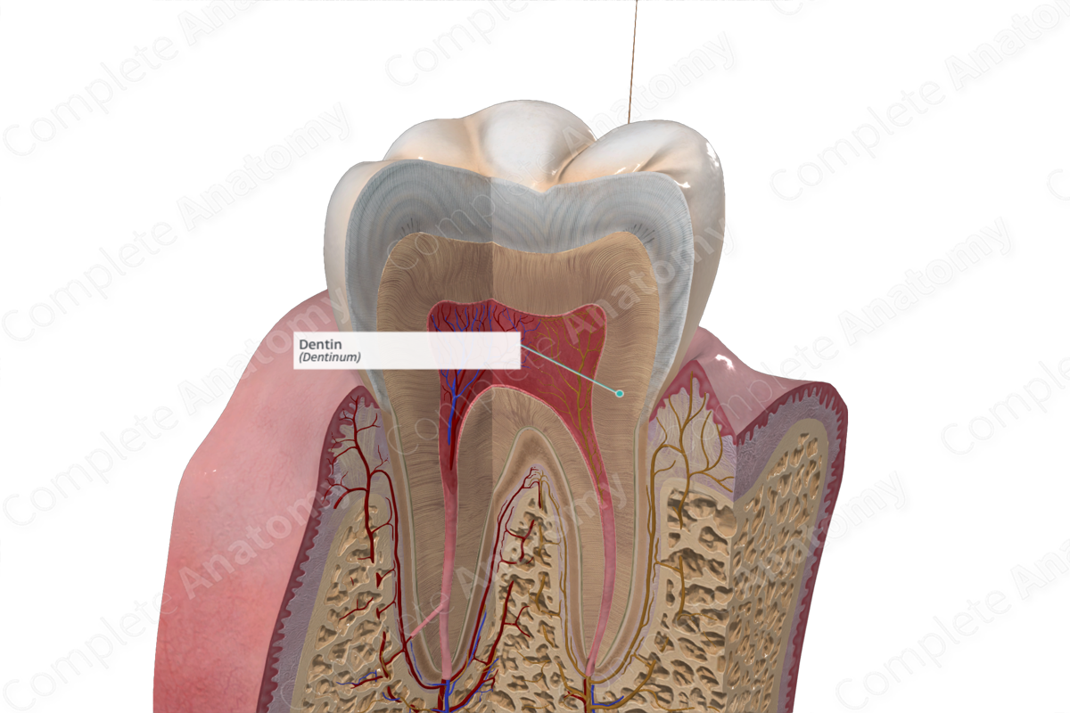
The dentin is the hard, bone-like portion of the tooth surrounding the pulp, covered by enamel on the crown and cementum on the root; it is harder and denser than bone but softer than enamel (Dorland, 2011).
Structure and/or Key Feature(s)
Dentin is the hard, light-yellow, avascular tissue which lies directly under the enamel and cement. Dentin constitutes the bulk of the tooth, with a mineral content of 70% dry weight, 20% organic matrix, and 10% water (Standring, 2016; Kerr, 2007).
It is produced by the odontoblasts located at the periphery of the dental pulp cavity through a process called dentinogenesis. Odontoblasts initially produce predentin which matures into dentin (Vishwakarma et al., 2014).
The inorganic mineral content of dentin largely consists of crystalline hydroxyapatite, smaller than those found in enamel, with the incorporation of some minor elements including carbonate, sodium, and magnesium. The organic matrix shares a similar composition to bone, consisting of collagen, non-collagenous proteins, phospholipids, and growth factors. Collagen constitutes 90% of the organic matrix, primarily type I collagen fibers, with the remaining 10% being non-collagenous proteins (Vishwakarma et al., 2014).
Structurally, dentin is comprised of many tubules which span its entire thickness. These tubules form as a result of the mineralization mechanism of the odontoblast cells and are arranged in a radial pattern around the pulp.
Three types of dentin exist: primary dentin, secondary dentin, and tertiary dentin.
—Primary dentin is defined as all dentin formed prior to root formation or completion. It is the most prominent dentin in the tooth and lies between the enamel and the pulp chamber.
—Secondary dentin is defined as all dentin produced after root completion. This is a slower and more intermittent deposition of dentin, which continues throughout life. It results in a reduction of the pulp chamber size and is distinguished from primary dentin by the random arrangement and reduction of dentinal tubules.
—Tertiary dentin is a reparative dentin which is produced in response to adverse external stimuli, e.g., advancing caries or tooth breakage. This dentin is characterized by poorly mineralized dead tracts, since odontoblasts in the affected region die and the tubules remain empty (Standring, 2016).
Different regions exist within the dentin, which are identified and recognizable by structural differences. Mantle dentin exists in the outermost dentin zone in the crown of the tooth and is characterized by the orientation of its collagen fibers, which are positioned perpendicular to the dentinoenamel junction. The peripheral dentin zone in the root of the tooth contains both a granular and hyaline layer. The latter lacks a tubular structure and functions to produce a strong bond between the dentin and cement (Vishwakarma et al., 2014; Kerr, 2007).
Unlike enamel, dentin is produced slowly throughout life; therefore it possesses an unmineralized zone, called predentin. This innermost layer of dentin is characterized by the presence of odontoblastic processes which facilitates the secretion of matrix components (Standring, 2016).
Anatomical Relations
Dentin lies superficial to enamel and cement.
Function
Dentin is the primary structural component of the tooth. It supports the enamel in maintaining rigidity of the tooth and protects the pulp cavity.
List of Clinical Correlates
—Dental dysplasia
References
Dorland, W. (2011) Dorland's Illustrated Medical Dictionary. 32nd edn. Philadelphia, USA: Elsevier Saunders.
Kerr, J. B. (2007) Atlas of Functional Histology. Elsevier Science Health Science Division.
Standring, S. (2016) Gray's Anatomy: The Anatomical Basis of Clinical Practice. Gray's Anatomy Series 41 edn.: Elsevier Limited.
Vishwakarma, A., Sharpe, P., Shi, S. and Ramalingam, M. (2014) Stem Cell Biology and Tissue Engineering in Dental Sciences. Elsevier Science.
