Quick Facts
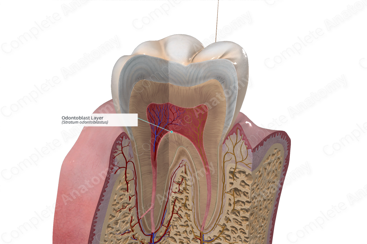
The odontoblast layer is the epithelioid odontoblastic zone, one to five layers thick, which forms the outer surface of the dental pulp adjacent to the dentin, resting on the subodontoblastic layer (Weil basal layer). It produces and maintains the dentin (Dorland, 2011)
Structure and/or Key Feature(s)
The odontoblast layer consists of odontoblasts cell bodies grouped in a row of 4 or 5 over a layer of Hoehl cells. Odontoblasts and Hoehl cells are derived from the neural crest (Hand and Frank, 2015).
Odontoblast cell bodies are large columnar cells that measure 3 µm wide and 20–40 µm long. Their cellular processes, also known as Tomes’ fibers, project across the predentin layer and penetrate dentinal tubules where they secrete peritubular dentin (also known as intratubular dentin). Odontoblasts have a polarized nucleus and well developed Golgi and rough endoplasmic reticulum.
Hoehl (Höhl) cells are described as the undifferentiated daughter cells of the mitotic process that remain immature due to their lack of contact with the basement membrane. The undifferentiated cells collectively become known as Hoehl’s cell layer. It is assumed that this cell layer differentiate into odontoblastic cells when original odontoblasts have died (Vishwakarma et al., 2014).
Anatomical Relations
The odontoblast layer is positioned subjacent to the predentin layer at the periphery of the pulp.
Function
The primary function of odontoblasts and the odontoblastic layer is dentin production. These cells are also thought to play a role in the transmission of sensory stimuli in the form of pain and have an additional role in the initiation of innate immune responses (Hand and Frank, 2015).
References
Dorland, W. (2011) Dorland's Illustrated Medical Dictionary. 32nd edn. Philadelphia, USA: Elsevier Saunders.
Hand, A. R. and Frank, M. E. (2015) Fundamentals of Oral Histology and Physiology. Wiley.
Vishwakarma, A., Sharpe, P., Shi, S. and Ramalingam, M. (2014) Stem Cell Biology and Tissue Engineering in Dental Sciences. Elsevier Science.
