Quick Facts
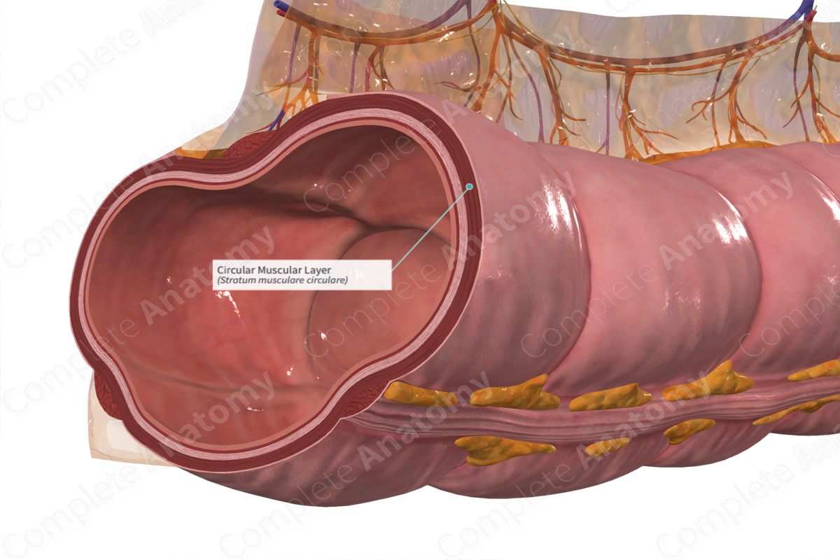
The circular muscular layer is the inner layer of circularly coursing fibers in the muscular coat (tunica muscularis) of the colon (Dorland, 2011).
Related parts of the anatomy
Structure/Morphology
The muscular layer of the colon is composed of smooth muscle organized into outer longitudinal and inner circular layers. These layers run perpendicular to each other. The inner circular layer extends the width of the colon and the outer longitudinal fibers are continuous along the length of the colon.
The circular layer of smooth muscle is thicker than the longitudinal layer between the teniae coli. It is not a uniform sheet of muscle and is divided by connective tissue clefts which penetrate the whole muscle and are indicated on the outer surface of the circular layer by superficial creases. Additionally, small fascicles of muscle pass between the circular and longitudinal layers, especially in the region of the teniae coli. This deviation of muscle fibers may help form the haustra (Standring, 2016).
The muscular layer receives its arterial blood supply from permeating capillaries coming from the submucosal arteriolar network and is innervated by the myenteric plexus.
Anatomical Relations
The circular layer is located between the submucosa and the outer longitudinal muscular layer. The myenteric plexus is located between the circular and longitudinal muscular layers.
Function
The structure of the circular layer impairs the luminal contents traveling backwards along the intestinal tract. It works with the longitudinal layer to provide peristaltic contractions, which facilitates the passage of the intestinal content to the anus for excretion.
References
Dorland, W. (2011) Dorland's Illustrated Medical Dictionary. 32nd edn. Philadelphia, USA: Elsevier Saunders.
Standring, S. (2016) Gray's Anatomy: The Anatomical Basis of Clinical Practice. Gray's Anatomy Series 41st edition edn.: Elsevier Limited.

